Download
1 / 20
220 likes | 348 Views
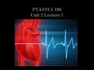
PTA 106 Unit 2 Lecture 1. Position of the heart and Associated Structures. Coronary trivia Pumps blood through 60,000 miles of blood vessels Pumps about 3,600 gal per day 2.6 million gal per year. Approximate Location of the heart projected to the surface. Landmarks

E N D
Position of the heart and Associated Structures • Coronary trivia Pumps blood through 60,000 miles of blood vessels • Pumps about 3,600 gal per day • 2.6 million gal per year
Approximate Location of the heart projected to the surface Landmarks • Superior R point: Is at the superior border of the R 3rd costal cartilage • Superior L point: Is located at the inferior border of the L 2nd costal cartilage • Inferior L point: (the apex) is located at of the heart in the L 5thintercostal space • Inferior R point: Is located at the superior border of the sixth R costal catilage
Physiology of Cardiac Muscle Contraction • Action potential initiated by the SA node • Action potential conducted to the purkinje fibers • Depolarization of sarcolemma opens voltage-gated fast Na+ channels causing rapid depolarization • Prolonged depolarization called the “plateau” involves opening of voltage-gated slow Ca2+ channels
Physiology of Cardiac Muscle Contraction • Repolarization is caused by opening of voltage-gated K+ channels • The prolonged depolarization causes an absolute refractory period where the cardiac muscle can not respond to additional stimulus.
The parts of an Electrocardiogram (EKG) during a cardiac cycle • P wave = atrial depolarization (Large P = atrial enlargement) • QRS complex = ventricular depolarization (Large Q = myocardial infarction) • T Wave = ventricular repolarization (Flat T = coronary artery disease) • P-Q interval = Time required for conduction from SA node to purkinje fibers
The parts of an Electrocardiogram (EKG) during a cardiac cycle • S-T segment = Time when ventricular myocardia is depolarized (elevated S-T indicates acute myocardial infraction} • Q-T interval= time form start of ventricular depolarization to ventricular repolarization. (Lengthened by myocardial damage)
The Cardiac Cycle: Atrial Systole Atrial Diastole Ventricular fillling Ventricular Ejection Ventricular Systole Ventricular Diastole Isovolumetric Contraction Isovolumetric Relaxation
The Cardiac Cycle: End-diastolic volume End-systolic volume
Cardiac Output (CO) • CO = volume of blood ejected from the left ventricle into the Aorta each minute. • CO = SV x HR • SV = stroke volume, volume of blood ejected from ventricle (70 ml) • HR = Heart rate, heartbeats per minute
Cardiac Output (CO) • Factors the effect SV 1. Preload: degree of stretch of the myocardium before contraction 2. Contractility: force of contraction of the ventricular myocardium 3. Afterload: Force or pressure that the ventricular myocardium must exceeded to open the semilunar valves.