Download
1 / 3

E N D
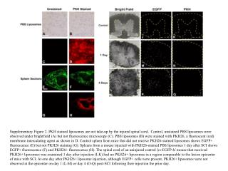
Supplementary Figure 2. PKH stained liposomes are not take-up by the injured spinal cord. Control, unstained PBS liposomes were observed under brightfield (A) but not fluorescence microscopy (C). PBS liposomes (B) were stained with PKH26, a fluorescent (red) membrane intercalating agent as shown in D. Control spleen from mice that did not receive PKH26-stained liposomes shows EGFP+ fluorescence (E) but not PKH26 staining (G). Spleens from a mouse injected with PKH26-stained PBS liposomes 1 day after SCI shows EGFP+ fluorescence (F) and PKH26+ fluorescence (H). The spinal cord of an uninjured control lys-EGFP-ki mouse that received PKH26+ liposomes was examined 1 day after injection (I-K) had no PKH26+ liposomes in a region comparable to the lesion epicenter of mice with SCI. At one day after PKH26+ liposome injection, although EGFP+ cells were present, PKH26+ liposomes were not observed at the epicenter on day 1 (L-M) or day 4 (O-Q) post-SCI following their injection the prior day.