Download
1 / 17
510 likes | 1.65k Views
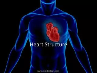
The Heart- Structure and Function. Lungs. Body cells. Our circulatory system is a double circulatory system. This means it has two parts. the right side of the system deals with deoxygenated blood. the left side of the system deals with oxygenated blood. The Heart.

E N D
Lungs Body cells Our circulatory system is a double circulatory system. This means it has two parts . the right side of the system deals with deoxygenated blood. the left side of the system deals with oxygenated blood.
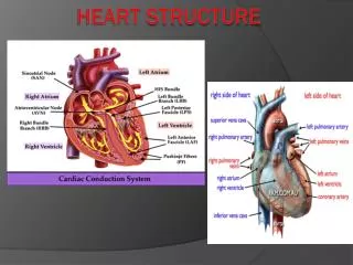
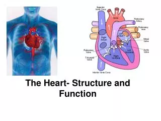
The Heart This is a vein. It brings blood from the body, except the lungs. These are arteries. They carry blood away from the heart. 2 atria Coronary arteries, the hearts own blood supply 2 ventricles The heart has four chambers now lets look inside the heart
The Heart The Heart Superior vena cava Left pulmonary arteries Aorta Left pulmonary veins Semilunar valve Left atrium Right atrium Atrioventricular/bicuspid / mitral valve Atrioventricular/ tricuspid valve Semilunar valve Inferior vena cava Left ventricle Right ventricle http://www.youtube.com/watch?v=D3ZDJgFDdk0
blood from the lungs blood from the body The heart beat begins when the heart muscles relaxand blood flows into all four chamber. The Heart Cycle and Sounds STEP ONE: Diastole Relax STEP TWO: Diastole Contract The atria then contract and the valves open to allow blood into the ventricles. This process results in a ‘lubb’ sound http://www.nhlbi.nih.gov/health/dci/Diseases/hhw/hhw_pumping.html
The valves close to stop blood • flowing backwards. • The ventricles contract forcing • the blood to leave the heart. • This process results in a ‘dubb’ sound. • At the same time, the atria are • relaxing and once again filling with • blood. STEP THREE: Systole The cycle then repeats itself. http://www.execulink.com/~ekimmel/forensic_flash_quiz4.swf
Co-ordination of the Cardiac Cycle • The heart is made of cardiac muscle. • When the cells receive an electricalimpulse they contract - causing a heartbeat. • Cardiac muscle can contract on its own, without needing nerveimpulses.
Sinoatrial node (SA node) This specialized excitatory muscle cells make an electric impulse. This signal travels across the atria causing them to contract and load the ventricles with blood.
Atrio-ventricular node (AV node) The AV node is a second bundle of excitatory muscle cells. Impulse from the SA Node to the AV node, causes an impulse in the AV node. Which causes the ventricles to contract and forces blood out of the heart to lungs and organs.
Electrocardiograph- see textbook pg 305, Fig 9-27 P to Q wave = SA impulse or atrial depolarization (loses electrical charge) Q→R → S wave = AV impulse or ventricular depolarization (loses electrical charge) S →T wave = ventricular repolarization (regains electrical charge) **Note: Atrial repolarization is NOT seen because it happens during the AV impulse** Records electrical activity of heart to monitor heart function.
Ventricular fibrillation is an abnormal heart rhythm that is disorganized and irregular.
Ventricular tachycardia is a rapid, regular heart rhythm that originates in the lower chambers of the heart.
Blood Pressure Is the pressure exerted on the walls of the arteries. There are 2 components to blood pressure: Systolic pressure is the pressure that the blood exerts on the aorta when blood leaves the heart during systole. Diastolic pressure is the pressure that the blood exerts on the aorta when noblood leaves the heart during diastole. Normal blood pressure is systolic = 120 mm Hg diastolic 80 mm Hg