Download
1 / 28
280 likes | 355 Views
The Heart In You. Produced by Jessica Owen Brittany Furches Julie Barrett. *. Heart Facts. Begins beating 4 weeks after conception Fully developed about 8 weeks after conception Force used to squeeze tennis ball 100,000 beats in a day 35 million beats in a year

E N D
The Heart In You Produced by Jessica Owen Brittany Furches Julie Barrett
* Heart Facts • Begins beating 4 weeks after conception • Fully developed about 8 weeks after conception • Force used to squeeze tennis ball • 100,000 beats in a day • 35 million beats in a year • 2.5 billion beats in a lifetime • Muscle force is twice as hard as person sprinting • Pumps about 1 million barrels of blood in lifetime • Heart performs enough work in one hour to lift 3,000 lbs. 1’ off the ground
Discovery of Circulation • William Harvey, 1628, theory of blood circulation first published • Showed that heart works like a pump • Described how blood flows from the heart to the lungs, back to the heart, out to the body, and back to the heart
Development of Heart Surgery • 1912, James B. Herrick, first diagnosis of a heart attack • 1938, Robert E. Gross performed first successful repair of a congenital heart defect. • 1952, Charles Hufnagel, operated on a beating heart & implanted first artificial heart valve • 1967, Christian Barnard, performed first human heart transplant • 1982, William DeVries, implanted first permanent artificial heart in human patient
* Stethoscope • 1816 (France) • Rene-Theophile-Hyacinthe Laennec • Wooden tube, • 1851, Arthur Leared • 1852, George Camman • 1961, Dr. David Littman • Open/closed chest-piece (appreciation/filter) • “two-sided chest-piece”, “combination”
* Stethoscope • One end is put on chest wall • Flat, thin side picks up vibrations of sounds coming through chest wall, picks up more high pitched sounds • “bell” side, shaped like a semi-circle, is rested very lightly on skin, picks up more low pitched sounds
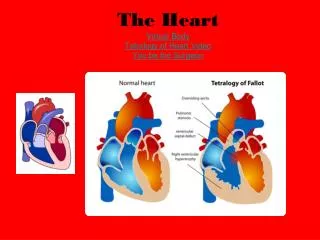
Right Coronary Artery Left Coronary Artery Superior Vena Cava Inferior Vena Cava Aorta Pulmonary Artery Pulmonary Vein Right Atrium Left Atrium Right Ventricle Left Ventricle Papillary Muscles Chordae Tendineae Tricuspid Valve Bicuspid Valve (Mitral) Pulmonary Valve Aortic Valve Parts of the Heart
Coronary Arteries-the vessels which bring blood to the heart muscle-Emerge from beginning of Aorta, near the top of the heart • Right Coronary Artery • Supplies back of the heart • Left Coronary Artery • Left Anterior Descending • Supplies front of heart • Left Circumflex • Wraps around left side and back of heart
Superior/Inferior Vena Cava • The mains veins which bring de-oxygenated blood from the body to the heart. • Veins from the head and upper body feed into the superior vena cava, which empties into the right atrium of the heart. • Veins from the legs and lower torso feed into the inferior vena cava, which empties into the right atrium of the heart.
Aorta • Largest single blood vessel in body • Approx. diameter of thumb, garden hose • Carries oxygen-rich blood from left ventricle to various parts of the body
Pulmonary Artery/Vein • Pulmonary Artery: The vessel that transports de-oxygenated blood from the right ventricle to the lungs • Pulmonary Vein: The vessel transporting oxygen-rich blood from the lungs to the left atrium
Left/Right Atrium • Left Atrium: receives oxygenated blood from the lungs through the pulmonary vein. • Right Atrium: receives de-oxygenated blood from the body through the superior vena cava & inferior vena cava
Left/Right Ventricle • Left Ventricle: receives oxygenated blood as the left atrium contracts. When the left ventricle contracts, the bicuspid valve closes & the aortic valve opens. • Right Ventricle: receives de-oxygenated blood from the body through the superior & inferior vena cava LEFT VENTRICLE RIGHTVENTRICLE
Papillary Muscles • Attach to lower portion of the interior wall of the ventricles • Connect to chordae tendineae • When papillary muscles contract, the valves open. When papillary muscles relax, the valves close.
Chordae Tendineae • “Heart Strings” • Tendons which link the papillary muscles to the tricuspid & bicuspid valve • As the papillary muscles contract & relax, the chordae tendineae transmit the resulting increase & decrease in tension to the valves, causing them to open/close
Tricuspid Valve • Separates the right atrium from the right ventricle • Closes as the right ventricle contracts preventing blood from returning to the right atrium, forcing it to exit the pulmonary valve into the pulmonary artery
Bicuspid Valve • Separates the left atrium from the left ventricle • Closes as the left ventricle contracts, preventing blood from returning to the left atrium, which forces it to exit through the aortic valve into the aorta
Pulmonary Valve • Separates the right ventricle from the pulmonary artery • As the ventricles contract, it opens to allow the de-oxygenated blood collected in the right ventricle to flow to the lungs • As the ventricles relax, it closes, preventing blood from returning to the heart
Aortic Valve • Separates the left ventricle from the aorta • Opens as ventricles contract to allow oxygenated blood in the left ventricle to flow throughout body • Closes as ventricles relax, preventing blood from returning to the heart.
* Pathway of Blood • Two pumps work simultaneously • Pulmonary Circulation • Pumps the blood to the lungs, where carbon dioxide is exchanged for oxygen • Systemic Circulation • Pumps the blood to the body, and releases the oxygen & nutrients
* Pulmonary Circulation Blood is Carried to the lungs De-oxygenated Blood Enters Heart
* Systemic Circulation Blood is pumped out to body Oxygen-rich blood comes back to heart
“Lub-dub” • “LUB” • Heard when valves between the upper chambers and lower chambers close • “DUB” • Heard when valves in the pulmonary and aortic arteries leaving the heart close, followed by a longer pause, when the heart relaxes to fill with blood for next beat
Common Heart Diseases • Coronary Artery Disease • (CAD) = narrowing of the arteries that supply blood to the heart, reducing amount of blood the heart muscle receives (“angina”) • Most common form of heart disease • Affects more men than women • Affects elderly more than younger people • May be hereditary • Risk Factors (controllable) • High BP, cigarette smoking, extreme obesity, stress
Common Heart Diseases • Heart Attack (myocardial infarction) • Death of heart muscle due to loss of blood supply • Almost always caused by blood clot on a cholesterol plaque in coronary artery • Pain: cardinal symptom • Tightness, heaviness in chest, frequently radiates to left arm or jaw • Risk Factors: • Smoking, high BP, elevated cholesterol, diabetes
Common Heart Diseases • Atherosclerosis • Formation of fat deposits on inner lining of arteries • Usually no symptoms until one or more arteries is so clogged with plaque that blood flow is severely reduced (“ischemia”) • Arteriosclerosis • Hardening, thickening, and loss of elasticity in artery walls • Symptoms: circulatory disturbances, skin temp. & color changes, altered pulse, headaches, dizziness, memory defects