Download
1 / 44
440 likes | 518 Views
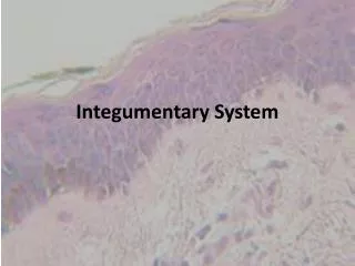
The Integumentary system. Integumentary system (skin). Skin: roles. 1- Protects against infection, mechanical shock, chemical injury 2- Prevents fluid loss 3- Sensor for touch, pain, temperature 4- Excretion of sweat 5- Immunity 6- Synthesis of vitamin D 7- Thermoregulation.

E N D
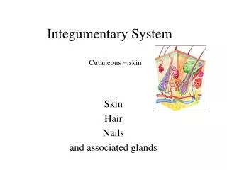
The Integumentary system Integumentary system (skin)
Skin: roles • 1- Protects against infection, mechanical shock, chemical injury • 2- Prevents fluid loss • 3- Sensor for touch, pain, temperature • 4- Excretion of sweat • 5- Immunity • 6- Synthesis of vitamin D • 7- Thermoregulation
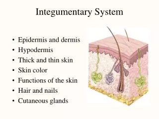
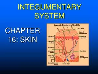
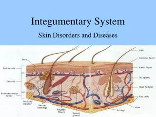
Integumentary system (skin) • Three layers: • Epidermis: the outer/superficial protective layer • Dermis: Deepest and thickest layer of the integumentary system, comprising 2 layers, the papillary and reticular layers - The “hypodermis” or subcutaneous tissue layer binds the dermis of the skin to the underlying muscle
Epidermis Several layers: -The main layer is located at the base • formed by the active, dividing cells or keratinocytes • -The keratinocytes multiply and push up, toward the surface of the skin • As they move away from the base and the blood supply, they die off, dry • Left are dried cells rich in keratin, a stringy protein • They form the upper layer, rich in keratin or corn • This keratin (corn) layer (in addition to an oily secretion) has a number of roles: • Keratin is somewhat water repellent (water proofs your skin)
Skin color Derived from 3 skin pigments: • Melanin • Carotene • Hemoglobin • Melanin (brown/black) – brown pigment secreted by the melanocytes. Stimulated by UV exposure to protect the basal layer from UV damages. Increased exposure lead to increased melanin synthesis • African American and Caucasian have the same number of melanocyte. However, the amount of melanin secreted varies with the human origin and the exposure to UV
Integumentary system (skin) Note: difference between the melanized cells of the stratum basale between the two skin tones. Also recall how the stratum corneum is transparent.
Skin color • Excess melanin: • Freckles = patches of dense melanin • “liver spots” = sun spots or age spots – excess melanocyte activity • seborrheic hyperkeratosis excess growth of keratinocytes
Skin color • Carotene: yellow pigment found in food (carrot) • Hemoglobin – red pigment found in the blood • Lack of oxygen (Hb only) turns the skin blue (cyanosis)
Integumentary system (skin) • Below the epidermis = Dermis • Variations in the layers forms valleys and grooves, seen on the surface of the skin as fingerprint. • The collagen fibers are distributed along a certain orientation surgeons tries to cut along the fibers, not across them, to minimize scarring
The dermis • Contains: • blood vessels • nerve fibers • Touch, pressure, temperature sensors • Muscle, the arrector pili • glands
Hypodermis • Below the dermis • Subcutaneous tissue (not actually part of the skin). • Can be thick, if rich in adipose tissue • Tthickness varies with location and individuals
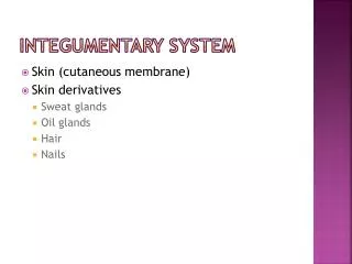
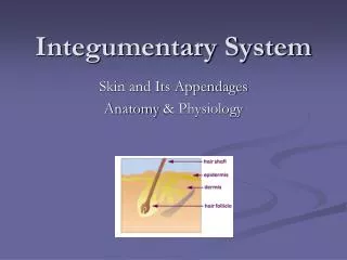
Epidermal derivatives: • Even though these structures are mostly located in the dermis they originate from the epidermis: • Hair • Nail • Glands - sebaceous glands - sweat glands - apocrine glands - eccrine glands
Epidermal derivatives: Hair • Each hair made of a shaft, root and bulb • Shaft is what you see (dead cells) • Root is below the skin • Bulb located at the base of the root within the follicle • Follicle is the sheath of epidermal cells around the root • Cells divide in the bulb, push their way up • This is how hair grows in length…cells growing • Roughly 1mm every 3 days • Speed of growth varies with individual and hair location
Hair Note how the stratum basale of the epidermis lines the hair follicle.
Hair • Defined lifespan: 3-4 months for eyelash, 3-4 years for scalp • Each hair is replaced by a new hair that pushes the old hair shaft out from the follicle • You have the same number of follicles…new hair just “reboots” the hair bulb to form a new unit. • In-grown hair often results when the new hair shaft cannot leave the follicle (due to restriction/constriction of the root). • Hirsutism: excessive body hair Hypertrichosis
Hair color • Hair color is define by the type and amount of pigment made in the stratum basale • Melanin (more = dark) • Red hair = presence of iron (trichosiderin) • Grey-white hair = lack of pigment due to dying melanocytes (and presence of air bubbles in the shaft) • Hair shape - round straight hair, oval curly hair • Hair thickness • - Depends on the size of the follicle and root
Nails • Composed of a compressed layer of stratum corneum cells • Hardness derived from dense keratin deposits. • Sometimes, the edge get stuck under the skin ingrown nail
Sebaceous glands • Derived from the epidermal cells • Open in the hair shaft • Secrete an oily substance, the sebum, making the skin water repellent • Accumulation of sebaceous secretion in a gland lead to the formation of a black head • If the gland becomes infected acne
Sudoriferous glands: sweat glands • Eccrine sudoriferous gland • Distributed all over the body (sweaty palms, back, chest etc.) • Non-smelly • Open on the skin • Apocrine sudoriferous gland • Armpits (axillary) and pubic regions – open into a hair shaft • Smelly sweat • Open into the hair shaft
Sudoriferous glands • Ceruminous glands: ear wax • Only located in the outer auditory canal • Cerumen = ear wax • For protection of the auditory canal from pathogenic invasion, • Also to lubricate the tympanic membrane.
Dermal structures • Arrector pili: • small muscle attached to hair root and base of epidermis • When pulled hair shaft stands up goose bumps • Vascular supply • nourish epidermis, hair root and dermis itself
Dermal structures: Sensory endings • Touch, pressure - sense touch and pressure • Temperature receptors • sense heat and cold • Pain receptors • Sense tissue damages
Clinical considerations • Wounds • Burns • Skin cancer • Aging
Wounds • Open skin is an entry door for bacteria risk of infection. • Gravity depends on depth and area involved. • Phases of healing: • Clot formation scab • Inflammatory response • Fibroblasts multiply granulations • Macrophages phagocytize debris • When dermis has filled up, epidermis can grow to cover the area • If severe wound: scar tissue
Inflammation • 4 cardinal symptoms • Bacteria in the wound make contact with defense cells such as mast cells mast cells release histamine • Histamine promotes increased permeability of blood vessels tissue swelling • Tissue swelling Pain
IInflammation • Symptoms of inflammation: • Redness • heat • Swelling • - pain • Bacteria also attract macrophages which release chemical promoting dilation of the capillaries (=vasodilation) more blood skin area becomes red (redness) and hot (heat)
Burns • Gravity of burns is determined by surface, depth and location • Surface: law of 9
Burns • Gravity of burns is determined by • 1) surface, 2) depth and 3) location • Depth: • First degree burn: involves epidermis only redness (erythema) – sun burn - painful - Skin heals and peals within 10 days no scarring
Burns • Depth: • Second degree burn: upper dermis involved blister - painful • Epidermis heals within few days little/no scarring
Burns • Depth: • Third degree burn: involves epidermis and entire dermis (and sometimes more) • Not painful! Why?
Skin tumors (benign and not) • Warts: due to a virus., treated by cryosurgery • Skin cancers • Basal cell carcinoma: most common, due to UV exposure, arises from basal cells, easily treated • Squamous cell carcinoma: from cells above basal cells, more invasive • Malignant melanoma: Due to melanocytes – changing moles – very invasive
Skin tumors (benign and not) • Skin cancers • Basal cell carcinoma: most common, due to UV exposure, arises from basal cell,,easily treated • Squamous cell carcinoma: from cells above basal cells, more invasive • Malignant melanoma: Due to melanocytes – changing moles – very invasive
Melanomas • Usually, starts from a mole • Watch for changes in shape, height or color of the mole • Melanomas are one of the deadliest cancers
Aging • Decrease in sebum secretion dry skin • Decrease in sweat gland secretion difficulties to cope with heat • Decrease in elastin fibers wrinkles • Decrease in adipose tissue in the dermis difficulties to cope with cold