Download
1 / 36
440 likes | 863 Views
Dr Taha Sadig Ahmed , MBBS , PhD ( England ) . Consultant , Clinical Neurophysiology . Associate Professor , Physiology Department , College of Medicine . Cerebellum ( Latin : Little Brain ). The cerebellum is the largest part of the hindbrain Relations :

E N D
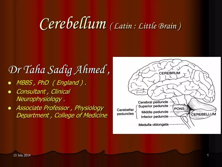
Dr Taha Sadig Ahmed , MBBS , PhD ( England ) . Consultant , Clinical Neurophysiology . Associate Professor , Physiology Department , College of Medicine Cerebellum ( Latin : Little Brain )
The cerebellum is the largest part of the hindbrain Relations : It is s located in the posterior cranial fossa , & has the following relations : Anteriorly: 4th ventricle, pons, and medulla oblongata Superiorly: it is covered by tentorium cerebelliI Inferiority: occipital bone It consists of 2 cerebral hemispheres which are interconnected by the vermis in the center. Surface shows parallel running folds known as Folia. Physiologic Anatomy (1) 4th Ventricle
Physiologic Anatomy (2) • The cerebellum influences movement on the ipsilateral side of the body. • Although it weighs only 10 % as much as the cerebral cortex , its surface area is about 75 % of that of the cerebral cortex . • It is connected to the brainstem on each side by the : • (1) Superior Peduncle has main connections to the Cerebrum . • (2) Middle peduncle has main connections to the Pons . • (3) Inferior Peduncle has main connections to the Medulla Oblongata .
Cerebellar Peduncles : Carry afferents from where ? Inputs to the Cereellum from the Cerebrum SuperiorCerebellar Peduncle Middle Cerebellar Peduncle Inputs to the Cerebellum from from the Pons Inferior Cerebellar Peduncle Inputs to the Cerebellum from the Medulla Oblongata
Corpus Cerebelli ( main body of cerebellum ) is divided into Anterior and Posterior lobe by Primary fissure. Floculonodular Lobe lies behind the posterolateral fissure Two cerebellar hemisphere are interconnected by the vermis. Anatomical Divisions of the Cerebellum
Anatomical Divisions of the , & Flocculonodular Lobe Cerebellum : Anterior Lobe , Posterior Lobe • The Primary Fissure divides the Corpus Cerebelli ( main body of cerebellum ) into Anterior and Posterior lobes. • The Floculonodular Lobe lies behind the posterolateral fissure • Thw two cerebellar hemisphere are interconnected by the vermis.
Spinocerebellum ( medial parts of hemispheres+ Vermis ) Neocerebellum (Lateral parts of hemispheres ) Hemisphere Posterolateral Fissure Flocculonodular Lobe Physiologic ( Functional ) divisions of the Cerebellum Neocerebellum , Spinocerebellum snd Vestibulocerebellum
Neocerebellum ( Posterior lobe ) Comprises the lateral portions of cerebellar hemispheres. Is newest from a phylogenetic point of view . It interacts with motor cortex in planning & programming of movements. The Neocerebellum is involved , in conjunction of the cerebral cortex , in planning & execution of skilled movements. It coordinate movements particularly of the distal limb muscles ( e.g., hand ) which are employed in skilful movement . NB the vemis projects to the brainstem & control the movement of axial and proximal limb muscle. Functionally , the Cerebellum is divided into 3 parts : Neocerebellum , Spinocerebellum , & Vestibulocerebellum Anterior Lobe Spino- cerebellum Vestibulocerebellum
Spinocerebellum ( Paleocerebellum) Consist of vermis & medial parts of the cerebellar hemispheres . It receives (1) Proprioceptive inputs ( afferents ) from all the body : Hence It is concerned with regulation of muscle tone . and it also receives (2) a copy of the “ Motor Plan “”from the motor cortex Therefore , by comparing plan with performance , it acts as a “ comparator “, and sends impulses back to the cortex to correct movement thereby it ccordinates & smoothes ongoing body movements • The vermis projects to the brainstem areas concerned with control of axial and proximal limb movements . • NB : Whereas the Neocerebellum controls particularly distal limb muscles that are neede for skilled movements , the vemis controls movement of axial and proximal limb muscle which are mainly concerned with gross postural adjustments . Anterior Lobe Spino- cerebellum Vestibulocerebellum
Vestibulocerebellum ( Floculonodular Lobe): Phylogenetically , it is the oldest part of the cerebellum ( hence it is also called Archicerebellum ) It has connections to the vestibular nuclei , consequently , it is concerned with balance & equilibrium And can induce changes in the VOR ( Vestibulocular Reflex ) Anterior Lobe Spino- cerebellum Vestibulocerebellum
The cerebellum has grey matter areas comprising the cerebellar cortex and the deep cerebellar nuclei ( DCN) . They are separated from each other by white matter ( nerve fibers ). The deep cerebellar nuclei are 4 in number , & are called the: (1) Dentate , (2) Globose , (3) Emboliform , & (4) Fastigial nuclei . Mossy Fibers , which are the primary afferents to the cerebellum , send collaterals to the deep nuclei and then proceed ( pass on ) to the cortex Cerebellar Organization
Cerebellar Cortex Efferenrts to the Deep Cerebellar Nuclei • Neocerebellar Cortex : • Projects ( sends its efferents ) to the Dentate Nucleus & from there to the Ventrolateral Nucleus of the Thalamus . • Spinocerebellar Cortex : • The Vermis projects to the Fastigial Nucleus & from there to the brainstem nuclei . • The hemispheric portions of the Spinocerebellum ( i.e., medial parts of the cerebellar hemispheres ) project to the Emboliform and Globose nuclei & from there to the brainstem nuclei . • Vestibulocerebellar Cortex : • Its efferents pass directly to the brainstem ( & not via the DCN) to regulate balance , equilibrium & the VOR ). • Consequently , the Deep Cerebellar Nuclei provide the only output of the Neocerebellum and Spinocerebellum .
The cerebellar cortex is made of layers (1) External Molecular layer , ( 2) Middle Purkinje Cell layer that is only one cell thick , & (3) Internal Granular layer Layers of the Cerebellar Cortex
The cerebellar cortex contains 5 types of neurons : Purkinje , Granule , Basket , Stellate & Golgi cells . (1) Purkinje Cells : Are amongst the biggest neurons in the body . Have very extensive dendritic arbors that extend throughout the Molecular Layer . Their axons , which are the only output from the cerebellar cortex , pass to the deep nuclei . Cells of the Cerebellar Cortex
Their cell-bodies are situated in the Granular layer . They receive input from the Mossy fibers and innervate the Purkinje cells . Each sends an axon to the Molecular layer , where the axon bifurcates to form a T . Because the branches of this “ T ” are straight and run for long distances , they are called Parallel Fibers . (2) Granule Cells( Origin of Parallel Fibers ) Granule cells
Because the dendrites of Purkinje cells are oriented at right angles to the Parallel fibers ( which are , actually , axons of Granule cells ) Each parallel fiber makes synaptic contacts with the dendrites of many Purkinje cells , Granule Cells ( continued ) Granule cells
And thus the parallel fibers and Purkinje cell dendritic trees form a grid of remarkably regular proportions
Trees of the • The other 3 types of neurons in the cerebellar cortex are inhibitory neurons : (3) Basket cells ( inhibitory to Purkinje ): • Are located in the Molecular layer • They are excited by Parallel fibers of Granule cells , & their output inhibits Purkinje cell discharge by a process of Feed-Forward Inhibition . • Their axons form a basket around the cell-body and axon hillock of each Purkinje cell they innervate . (4) Stellate cells ( inhibitory to Purkinje ): • Similar to Basket cells they are excited by Parallel fibers of Granule cells , & their output inhibits Purkinje cell discharge by a process of Feed-Forward Inhibition . • They differ from Basket cells only in being more superficially located in the cortex than Basket cells .
Golgi cells are located in the Granular layer . Their dendrites , which project into the Molecular layer , receive inputs from the Parallel fibers . Their cell bodies receive input via collateralsfrom the incoming Mossy fibers and the Purkinje cells Their axons project to inhibit the dendrites of the Granule cells . They are excited by (1) Mossy fibers (2) Purkinje cells , & (3) Parallel fibers ( of Granule cells ). They inhibit the excitatory action of Mossy fibers on Granule cells . (5) Golgi cells
There are 2 main inputs to the cerebellar cortex : the Clombing Fibers and Mossy Fibers , both of which are excitatory . Climbing Fibers : The climbing fibers come solely from the Inferior Olivary Nucleues They provide an indirect proprioceptive input to the cerebellar cortex bringing to it proprioceptive information from all parts of the body via relays in the Inferior Olive (which receives proprioceptive inputs from all over the body parts ) Each climbing fiber projects to the dendrites of Purkinje cells , around which it entwines like a climbing plant . The Main Inputs (Afferents ) to the Cerebellar Cortex (1)
Mossy Fibers : (1) These , unlike Climbing Fibers ( which provide an indirect proprioceptive input ) do provide a direct proprioceptive pathway( input ) to the cerebellar cortex , from all parts of the body , and , in addition (2) Provide inputs from the Motor Area ( M1) & related areas of the Cerebral Cortex( indirectly , via relays in the pontine nuclei ). They end on the dendrites of Granule cells in complex synaptic groupings called Glomeruli . The Glomeruli also contain the inhibitory endings of the Golgi cells. The Main Inputs (Afferents ) to the Cerebellar Cortex (2) Climbing Fibers Mossy Fibers
Corollary ( summary ) of effects of different cells & afferents on Purkinje cells • (A) Excitatory • The fundamental circuits of cerebellar cortex are thus relatively simple : (1) climbing fiber inputs exert a strong excitatory effect on single Purkinje cells , whereas (2) Mossy fiberinputs exert aweak excitatoryeffect onmanyPurkinje cells via the Granule cells . • (B) Inhibitory (1) Basket cells (2) Stellate cells (3) Golgi cells • Golgi cells are excited by (1) Mossy fibers (2) Purkinje cells , & (3) Parallel fibers . • They inhibit the action of Mossy fibers on Granule cells Both are excited by Parallel fibers of Granule cells , & their output inhibits Purkinje cells ( Feed-Forward Inhibition ) .
Q : What are the Neurotransmitters Secreted by in the Cerebellar Cortex Neurons ? • Purkinje cells • Basket cells • Stellate cells • Golgi cells • Granule cells Glutamate Secrete GABA
Functional Significance of Cerebellar Cortex Circuitry • The DCN are excitatory to the Brainstem nuclei & Thalamus . • The circuitry of the Cerebellar Cortex seems to be solely concerned with modulating the • (1) Timing • (2) strength • Remember that : The DCN are excited by both Mossy & Climbing fibers , but are inhibited by Purjkinje cells . • Hence activity in Mossy & Climbing fibers excite the DCN . • But these are also excitatory to Purkinje cells which inhibit the DCN . • Thus the effect of the afferent inputs seems to activate the DCN initially , & then , after a latency of time ( of a few ms perhaps ) , to switch them off via exciting the Purkinje cells ( remember that more synapses mean more latency ) . of the excitatory action of the DCN on the Brainstem & Thalamus
The Primary Afferents that Converge to Form the Mossy Fiber or Climbing Fiber Inputs to the Cerebellum,
Remember • Cerebellum hemispheres control the same ( ipsilateral ) side of the body . • Purkinje cells are the main output neurons of the cerebellar cortex & project to the deep nuclei of the cerebellum. • They are inhibitory to the DCN . • The deep cerebellar nuclei ( DCN ) project out to brainstem and thalamic targets via the superior cerebellar peduncles. They are excitatory , but in turn , are themselves inhibited ( switched off ) by Purkinje cells . • Flocculonodular lobe is important for regulation of balance , equilibrium & the VOR .
Cerebellar Hemispheric Lesions • Cerebellar lesions cause no paralysis or sensory deficit . • When not moving , there are no externally obvious signs . • However , upon physical examination , signs such as hypotonia and pendular reflexes can be elicited . • Once the patients attempts movement , ataxia appears . • What is ataxia ? Ataxia is incoordination of due to errors in the rate , range , force and direction of movement . • With circumscribed lesions , the ataxia may be confined/localized to only one part of the body .
The Difference Between Lesions of the Cerebellar Cortex & Lesions of DCN • If only the cortex of the cerebellum is involved , the movement abnormalities gradually disappear as “ compensation ” occurs . • However , lesions of the DCN produce more generalized defects , and abnormalities are permanent . • For this reason , care should be taken to avoid damaging the DCN when surgery is undertaken to remove a tumor involving part of the cerebellar cortex .
A/ Hemispheric Lesions • I/Ataxia (lack of coordination of muscle movement ) , which is manifested by • (1) Wide-based , unsteady “ drunken , or staggering “ gait . • (2) Scanning speech • (3) Dysmetria( also called Past-Pointing ) : attempting to touch an object with a finger results in overstretching to one side or the other this promptly initiates a gross correction action ( corrective action ) , but the correction overshoots to the other side Consequently , the finger oscillates back and forth . • This oscillation is the (4) “ Intention Tremor ” , which is characteristic of cerebellar disease . • This cerebellar tremor , unlike that of Parkinson’s disease , is absent at rest .
Hemispheric Lesions ( Contd ) • II/ Inability to “ put on the brakes ” i.e., inability to stop movement promptly . Normally , for example , flexion of the forearm against resistance is quickly checked when the resistance force is suddenly broken off . The patient with cerebellar disease can not break the movement of the limb , and the forearm flies back in a wide arc . This abnormal response is known as the “ Rebound Phenomenon ”. • III/ Adiadochkinesia ( Dysdiadochkinesia ) : Inability to perform rapidly alternating opposite movements such as repeated pronation and supination of the hands . • IV/ Difficulty in performing actions that involve simultaneous motions at more than one joint . The patient dissects such movements and carries them out one joint at a time , a phenomenon known as “ Decomposition of Movement ” .
B/ Flocculonodular Lobe Lesions • Midline cerebellar tumors in children , arising from the “ Nodule ” , early in their course (& before affecting the rest of the cerebellum) , damage first the Flocculonodular lobe . • Such a child is afraid ( & reluctant ) to stand erect and move without support . • This is because if he tries to walk , he does so in a staggering fashion on a broad base , & tends to fall . • Moreover , selective Flocculonodular lobe lesions may cause vertigo
Role of the Cerebellum in Learning (1) • The cerebellum is concerned with learned adjustments that make coordination easier when a given task is performed over & over • As a motor task is learned , activity in the brain shifts from the Prefrontal ( cerebral ) Cortex to the (1) Parietal Cortex , (2) M1 , & (3) Cerebellum . • The basis of learning in the cerebellum is the input via the Olivary Nucleus. • It is worth noting , in this regard , that each Purkinje cell receives inputs from 250,000 to 1,000,000 Mossy fibers. • By contrast , each Purkinje cell receives only a single ( only one ) Climbing fiber from the inferior olive, and this fiber makes 200-3000 synapses on the Purkinje cell .