Download

1 / 90
900 likes | 1.16k Views
Keys to 2402 Models. Photographs and Keys by Jeff Beck Department of Math and Natural Sciences Collin County Community College. 1. Frontal bone (Os frontale) 2. Temporal bone ( Os temporale) 3. Sphenoid bone (Os sphenoidale) 4. Occipital bone (Os occipitale)

E N D
Keys to 2402 Models Photographs and Keys by Jeff Beck Department of Math and Natural Sciences Collin County Community College
1. Frontal bone (Os frontale) 2. Temporal bone ( Os temporale) 3. Sphenoid bone (Os sphenoidale) 4. Occipital bone (Os occipitale) 5. Parietal bone (Os parietale) 6. Ethmoid bone (Os ethmoidale) 7. Maxilla (upper jaw) 8. Zygomatic bone (Os zygomaticum) 9. Palatine bone (Os palatinum) 10. Lacrimal bone (Os lacrimale) 11. Inferior nasal concha (Concha nasalis inferior) 12. Nasal bone (Os nasale) 13. Vomer 14. Mandible (lower jaw) 15. – 21. Cervical vertebrae
Endocrine glands and their parts • Pituitary gland or hypophysis • Adenohypophysis • Neurohypophysis • Infundibulum • Thyroid gland • Lateral lobe of thyroid • Isthmus of thyroid • Parathyroid gland • Suprarenal or adrenal gland • Capsule of suprarenal gland • Cortex of suprarenal gland • Medulla of suprarenal gland • Pancreatic islets of Langerhans • Alpha cells of pancreatic islets • Beta cells if pancreatic islets • Testis or testicle (male) • Seminiferous tubules • Epididymis • Ductus deferens • Ovary (female) • Blood vessels to ovary • Medulla of ovary • Developing follicles in ovary • Corpus luteum • Body organs and structures closely • associated with endocrine glands • 25. Diencephalon • Trachea • Carotid artery • Arch of aorta • Heart • Kidney • Pancreas • Uterus • Oviduct or fallopian tube
BRAIN • End brain • Interbrain • Midbrain • Afterbrain • Marrowbrain • I, II, III Cerebrum • Superior longitudinal sinus • Tentorium cerebelli • Little brain • a. arbor vitae cerebelli • Trabs cerebri • a. rostrum corporis callosi • b. genu corporis callosi • Septum pellucidum • Fornix • Anterior commissure • Middle commissure • Posterior commissure • Thalamus • Foramen of Monro • Aqueduct of the cerebrum • Ventriculus quartus • Corpus pineale • Chiasma opticum • Hypophysis • Corpus mamillare • Quadrigeminal plate • Upper velum palati • Bridge • Lengthening of spinal marrow • Spinal marrow
Hyoid bone • Larynx • 2. Thyroid cartilage • 3. Annular cartilage • 4. Epiglottis • 5. Arytenoid cartilage • 6. Thyrohyoid membrane • 7. Thyrohyoid muscle • 8. M. cricothyroideus • 9. M. arytaenoideus transverses • 10. M. arytaenoideus obliquus • 11. Posterior crycoarytenoid muscle • 12. Laryngeal pouch • 13. Superior laryngeal artery • 14. Inferior thyroid vein • B. Thyroid gland
Heart apex • Interventricular septum • Right auricle • Left auricle • Right auricular appendage • Left auricular appendage • Right ventricle • Left ventricle • Superior vena cava • Inferior vena cava • Tricuspid valve • Pulmonary artery • Pulmonary veins • Bicuspid valve • Aorta • Semilunar valves • Chordae tendinae 2. Papillary muscles 3. Ascending branch of the aorta 4. Right coronary artery 5. Left coronary artery 6. Great heart vein 7. Arch of the aorta
Aorta • Brachiocephalic artery • Left common carotid artery • Left subclavian artery • Superficial cardiac plexus • Left vagus nerve • Recurrent laryngeal nerve • Ligamentum arteriosus • Left pulmonary artery • Left bronchus • Left bronchial artery • Pulmonary nerve plexus • Intercostal arteries • Thoracic duct • Azygos vein • Esophagus • Right vagus nerve • Right bronchial artery • Trachea • Right main bronchus • Right pulmonary artery • Tracheobronchial lymph nodes • Right ventricle • Left ventricle • Left atrium • Right atrium • Right auricle • Conus arteriosus • Left auricle 30. Apex 47. Azygos vein 64. Crista supraventricularis 31. Pulmonary artery 48. Left pulmonary veins 65. Tricuspid valve, anterior cusp 32. Aorta 49. Right pulmonary veins 66. Tricuspid valve, posterior cusp 33. Pericardium 50. Marginal branch of right coronary artery 67. Tricuspid valve, medial cusp 34. Superior vena cava 51. Coronary sulcus 68. Anterior papillary muscle 35. Right coronary artery 52. Anterior interventricular sulcus 69. Posterior papillary muscle 36. Anterior cardiac veins 53. Crista terminalis 70. Septal papillary muscle 37. Left coronary artery 54. Sinoatrial node 71. Trabeculae carnae 38. Anterior interventricular artery 55. Atrioventricular node 72. Right branch of bundle of His 39. Circumflex artery 56. Opening of coronary sinus 73. Moderator band 40. Great cardiac vein 57. Limbus fossa ovalis 74. Pulmonary conus 41. Marginal branch of left coronary artery 58. Fossa ovalis 75. Pulmonary semilunar valve, posterior 42. Oblique vein of left atrium 59. Valvula venae cavae 76. Pulmonary semilunar valve, anterior 43. Coronary sinus 60. Musculus pectinatus 77. Pulmonary semilunar valve, anterior 44. Dorsal interventricular vein 61. Tricuspid valve anterior cusp 78. Foramen ovale 45. Left dorsal ventricular vein 62. Tricuspid valve medial cusp 79. Opening of pulmonary vein 46. Inferior vena cava 63. Tricuspid valve posterior cusp 80. Bicuspid valve, anterior cusp 81. Bicuspid valve, posterior cusp 82. Anterior papillary muscle 83. Chordae tendinae 84. Posterior papillary muscle 85. Trabecular carnae 86. Aortic semilunar valve, right anterior 87. Aortic semilunar valve, left anterior
The major parts of the circulatory system 15. Aorta 37. Tunica media • Heart 16. Inominate atery 38. Tunica externa • Circulatory system 17. Left common carotid • Vein 18. Subclavian artery • Artery 19. Epicardium • Principal structures of the heart 20. Myocardium • Muscle wall of right ventricle 21. Endocardium • Muscle wall of left ventricle • Intraventricular septum 22. Heart • Right atrium 23. Kidneys • Left atrial wall 27. Abdominal aorta • Papillary muscles 28. Femoral arteries • Tricuspid valve 29. Arteries • Bicuspid valve 30. Subclavian arteries • Pulmonary semilunar valve 31. Arteries • Aortic semilunar valve 32. Arteries • Superior vena cava 33. Wall of vein • Inferior vena cava 34. Valve in vein • Pulmonary artery 35. Typical artery • Pulmonary veins 36. Tunica interna of artery
Vena frontalis • Vena temporalis supervicialis • Superficial temporal artery • 3a. Maxillary artery • Occipital artery • Supratrochlear artery • Angular artery and vein • Facial artery and vein • 7a. Lingual artery • Retromandibular vein • Vena jugularis interna • Superior thyroid artery • Vertebral artery • Thyrocervical trunk • Costocervical trunk • Transverse scapular artery • Subclavian artery and vein • Superior vena cava • Common carotid artery • 18a. External carotid artery • Arch of aorta • Descending aorta • Axillary artery and vein • Vena cephalica • 22a. Anterior humeral circumflex artery • 22b. Posterior humeral circumflex artery • Thoracodorsal artery • Brachial artery and vein • Acromiothoracic artery • Left subclavian artery • Vena bascilica • Superior ulnar collateral artery • Ulnar artery • Common interosseous artery • Vena mediana antibrachii • Radial artery • Vena cephalica • Superficial palmar arch • Common volar digital arteries and veins • Artery profunda brachii • Radial recurrent artery • Dorsal carpal branchof the radial artery • Posterior branch of the interosseous artery • Dorsal metacarpal arteries • First dorsal metacarpal arteries 46. Vena pulmonalis 47. Pulmonary artery 48. Truncus pulmonalis 49. Lung (left) 50. Right atrium 51. Left atrium 52. Right ventricle 53. Left ventricle 54. Aortic valve 55. Pulmonary valve 56. Interventricular septum 57. Midriff 58. Liver 59. Venae hepaticae 60. Gastric veins, right and left 61. Common hepatic artery 61a. Truncus coeliacus 62. Superior mesenteric vein 63. Inferior vena cava 64. Renal artery and vein 65. Superior mesenteric artery 66. Lienal artery and vein 67. Left gastric artery 68. Aorta abdominalis 69. Inferior mesenteric artery 72. Common iliac artery and vein 73. External iliac artery and vein 74. Middle sacral artery 75. Internal iliac artery and vein 76. Ascending branch of the lateral femoral circumflex artery 77. Descending branch of the lateral femoral circumflex artery 78. Femoral artery 79. Deep femoral artery 80. Popliteal artery and vein 81. Posterior tibial artery 82. Anterior tibial artery 83. Arteria dorsalis pedis 84. Lateral plantar artery 85. Superficial artery 86. Vena profunda femoris 87. Medial femoral circumflex artery 88. External pudendal artery 89. Vena femoralis 91. Vena saphena parva 93. Perforating arteries 95. Medial superior genicular artery 95a. Lateral superior genicular artery 96. Lateral inferior genicular artery 96a. Medial inferior genicular artery 97.Highest genicular artery 98. Communicating perforating artery 99. Vena saphena magna 100. Rete venosum dorsale pedis 101. Arteria dorsalis pedis 102. Arcute artery 103. Arcus venosus dorsalis pedis 104. Dorsal metatarsal arteries 104a.Venae digitales pedis dorsales 106. Anterior tibial artery Human blood circulation model
Transverse sinus 23. Olfactory bulb • Straight sinus 24. Olfactory tract • Superior sagittal sinus 25. Optic nerve • Great vein of the cerebrum 26. Oculomotor nerve • 2. Sigmoid sinus 27. Trochlear nerve • 3. Superior petrous sinus 28. Trigeminal nerve • 4. Inferior petrous sinus 29. Trigeminal ganglion • 5. Cavernous sinus 30. Ophthalmic nerve • 6. Intercavernous sinuses 31. Maxillary nerve • 7. Occipital sinus 32. Mandibular nerve • 8. Internal carotid artery 33. Abducens nerve • 9. Ophthalmic artery 34. Intermediate nerve • 11. Posterior communicating artery 35. Facial nerve • 12. Posterior cerebral artery 36. Nervus vestibulocochlearis • 13. Basilar artery 37. Glossopharyngeal nerve • 14. Superior artery of the cerebellum 38. Vagus nerve • 15. Artery of the labyrinth 39. Accessory nerve • 16. Inferior anterior artery of the cerebellum 40. Hypoglossal nerve • 17. Vertebral artery 41. Cervical nerves • 18. Anterior spinal artery 42. Hypophysis • 19. Inferior posterior artery of the cerebellum • 20. Middle meningeal artery • 21. Diploic veins • 22. Diploe
The Human Brain • TELENCEPHALON • Frontal lobe 17. Choroid plexus 36. Pons • Parietal lobe 18. Hippocampus 37. Medulla oblongata • Occipital lobe Diencephalon 38. Olive • Temporal lobe 19. Thalamus 39. Pyramid • Central sulcus 20. Hypothalamic sulcus 40. first cervical nerve • Precentral gyrus 21. Hypothalamus Cervical Nerves • Postcentral gyrus 22. Interthalamic adhesion I. Olfactory • Olfactory bulb 23. Pineal body II. Optic • Anterior commissure 24. Left mamillary body III. Oculomotor • Corpus callosum 25. Pituitary gland IV. Trochlear • a. Genu 26. Choroid plexus V. Trigeminal • b. Trunk 27. Caudate nucleus VI. Abducens • c. Splenium 28. Pulvinar VII. Facial • d. Rostrum 29. Medial geniculate body VIII. Vestibulocochlear • 11. Septum pellucidum 30. Lateral geniculate body IX. Glossopharyngeal • 12. Fornix 31. Quadrigeminal lamina X. Vagus • 13. Posterior commissure 32. Tegmentum XI. Accessory • 14. Insula 33. Cerebral peduncle XII. Hypoglossal • 15. Internal capsule 34. Cerebral aquaduct Circle of Willis • 16. Lateral ventricle 35. Cerebellum
ARTERIES • Axillary • Humeral circumflex • Brachial • Brachial profunda • Superior ulnar collateral • Inferior ulnar collateral • Anterior ulnar recurrent • Radial • Ulnar • Anterior Interosseous • Posterior Interosseous • Deep palmar arch • Superficial Palmar arch • Middle collateral • Radial collateral • VEINS • 16. Median • 17. Basilic • 18. Axillary • 19. Median cubital • 20. Cephalic • 21. Accessory cephalic
Placenta • Fetal side of the placenta • Maternal side of the placenta, • with maternal veins • Fetal membrane • Umbilical cord • Umbilical vein • Umbilical arteries • Umbilical ligament • Liver • 9a. Ductus venosus Arantii • Heart • Portal vein • Lower vena cava • Right vestibule • Foramen ovale • Upper vena cava • Right ventricle • Tricuspid valve • Pulmonary artery • Ventricular septum • Left vestibule • Left ventricle 22. Bicuspid valve 23. Aortic arch 24. Ductus arteriosus Botalli 25. Descending aorta 26. Diaphragm 27. Thymus 28. Right lung 29. Left lung 30. Windpipe 31. Oesophagus 32. Stomach 33. Duodenum 34. Pancreas 35. Spleen 36. Kidney 37. Adrenal gland 38. Renal duct 39. Bladder 40. Womb 41. Ovary 42. Rectum
Occipital lymph nodes • Superficial cervical lymph nodes • Cubital lymph nodes • Deep cervical lymph nodes • Axillary lymph nodes • Right lymphatic duct • Tracheobronchial lymph nodes • Hepatic lymph nodes • Coeliac lymph nodes • Common iliac lymph nodes • Inguinal lymph nodes • Parotid gland lymph nodes • Submandibular lymph nodes • Submental lymph nodes • Jugular trunk • Thoracic duct • Subclavian trunk • Bronchial lymph nodes • Cisterna chyli • Gastric lymph nodes • Mesenteric lymph nodes • Right venous angle • Right lung • Liver • Gall bladder • Portal vein • Caecum with appendix vermiformis • Bifurcation of trachea • Stomach • Transverse colon
Nose • Nostril • Nasal cavity • Pharynx (nasal part) • Pharynx (oral part) • Pharynx (laryngeal part) • Upper lip • Lower lip • Hard palate • 9a. Uvula • Mandible • Teeth • Tongue • Oral cavity • Epiglottis • Larynx • Thyroid cartilage of larynx • Cricoid cartilage of thyroid • Vocal cords • Trachea • Left main bronchus • Bronchus • Bronchus • Right main bronchus • Bronchus • Bronchus • Left lung • Right lung • Rib cage • Diaphragm • Heart • Pulmonary arteries • Aortic arch • Superior vena cava • Bronchioles • Alveoli • Air sacs • Pulmonary venule • Pulmonary arteriole • Cerebrum • Cerebellum • Pons • Spinal cord • Frontal sinus • Sphenoid sinus • Cervical vertebrae • Pulmonary veins
Hyoid bone • Larynx • 2. Thyroid cartilage • 3. Annular cartilage • 4. Epiglottis • 5. Arytenoid cartilage • 6. Thyrohyoid membrane • 7. Thyrohyoid muscle • 8. M. cricothyroideus • 9. M. arytaenoideus transversus • 10. M. arytaenoideus obliquus • 11. Posterior crycoarytenoid muscle • 12. Laryngeal pouch • 13. Superior laryngeal artery • 14. Inferior thyroid vein • B. Thyroid gland c. Inferior lobe Left lung d. Superior lobe e. Inferior lobe • C. Windpipe • Bifurcation of the windpipe • Right principal bronchus • Right vena subclavia • 3a. Right subclavian artery • Right common carotid artery • Right vena jugularis interna • Right vena brachiocephalica • Trunk of brachiocephalica artery • Left pulmonary artery • D. Gullet • E. Lung • Right lung • Superior lobe • Middle lobe
Left half of the nasal cavity 18. Large anterior palatine nerve • Frontal bone with portion of frontal sinus 19. Ramilication of the large anterior palatine • Nasal bone nerve on the lower surface of the hard palate • Maxilla 20. Anterior or superficial branch • Palatine bone 21. Spheno-palatine artery and vein • Sphenoid bone with sphenoid sinus 22. Anterior nasal artery and vein • Superior nasal concha 23. Olfactory nerves, outer group • Middle nasal concha • Inferior nasal concha B. Right half of nasal cavity with septum • Upper nasal meatus 24. Nasal septum • Middle nasal meatus 25. Olfactory nerve, inner group • Inferior nasal meatus 26. Posterior nasal artery and vein • Opening of the Eustachian tube 27. Nasopalatine nerve • Foramen of the sphenoid sinus 28. Canal of Scarpa • Foramen of naso-lacrimal canal 29. Soft palate • Olfactory tract 30. Uvula • Olfactory bulb 31. Roof of mouth cavity • Spheno-palatine ganglion 32. Mucous lining of nasal and mouth cavity
Hyoid bone • Thyroid cartilage • Cricoid cartilage • Epiglottis • Arytenoid cartilages • Thyroid • Parathyroid glands
Hyoid bone • Thyroid cartilage • Cricoid cartilage • Epiglottis • Arytenoid cartilages • Corniculate cartilage • Trachea
Intra-pulmonary bronchi 21. Anastomosis between brachial artery • Non-cartilaginous bronchioles and pulmonary vein • Respiratory bronchioles 22. Alveolar capillary network • Alveoli 23. Anastomosis of an obstructed artery • Mucous membrane and pulmonary vein • Elastic fibers 24. Dense capillary network • Smooth muscle 25. Anastomosis between pulmonary artery • Pulmonary plexus and vein • Mucous glands of the bronchi • Fibro-cartilaginous layer • Pulmonary pleura • Limiting membranes • Dense capillaries • Collaginous fibers • Elastic fibers • Epithelium • Bronchial vein plexus • Pulmonary vein • Pulmonary artery • Bronchial artery
Hard palate • Soft palate • 2a. Uvula • Genioglossus muscle • Pharynx • Esophagus • Stomach • Cardiac orifice • Epigastric fossa • Lesser curvature • Greater curvature • Gastric canal • Pylorus • Duodenum • Longitudinal fold of the duodenum • Bile duct • Excretory duct of the pancreas • Gall bladder • Pancreas • Spleen • Liver • Right lobe • Left lobe • Quadrate lobe • Portal vein • Hepatic artery • Small intestine • Jejunum • Ileum • Ileo-cecal valve • Cecum • Vermiform appendix • Large intestine • Ascending colon • Transverse colon • Descending colon • Sigmoid flexure • Rectum • Nasal cavity • Larynx
Mouth • Teeth • Tongue • Parotid salivary gland • Submandibular and sublingual salivary glands • 6. Pharynx • 7. Esophagus • 8. Stomach • 9. Liver • 10. Gallbladder • 11. Duodenum • 12. Pancreas • 13. Jejunum • 14. Ileum • 15. Ileocecal valve • 16. Cecum • 17. Ascending colon • 18. Right colic flexure • 19. Transverse colon • 20. Left colic flexure • 21. Descendingcolon • 22. Sigmoid colon • 23. Rectum • 24. Vermiform appendix • 25. Fundus of stomach • 26. Longitudinal muscle layer of stomach • 27. Circular muscle layer of stomach • 28. Oblique muscle layer of stomach • 29. Tunica serosa • 30. Rugae of stomach • 31. Pylorus (sphincter muscle) • 32. Cystic duct • 33. Hepatic duct • 34. Common bile duct • 35. Interior wall intestinal fold • 36. Vascular network • 37. Intestinal lymph drainage • 38. Villi • 39. Villus of small intestine • 40. Lymphatic vessel • 41. Arteriole • 42. Venule • 43. Capillary network • 44. Secretory goblet cells
Central incisors 17. Crown of tooth (corona dentis) • Lateral incisors 18. Root of tooth (Radix dentis) • Canines or Cuspids 19. Enamel • First premolars (Bicuspids) 20. Dentine • Second premolars (Bicuspids) 21. Pulp cavity • First molars 22. Pulp canal • Second molars 23. Cementum • Third nolars 24. Apical foramen • Transverse palatine folds 25. Gum • Hard palate 26. Periodontal membrane • Soft palate 27. Rim of alveolus • Pharyngopalatine arch 28. Alveolar bone • Uvula 29. Haversian canal • Alveolar nerve 30. Bone of lower jaw • Alveolar artery • Vein
1. 2. 3. 4. 5. D 10/1 lower incisor tooth D 10/2 lower canine tooth D 10/3 lower premolar tooth D 10/4 lower molar tooth with caries D 10/5 upper molar tooth 1. crown of the tooth 2. dental neck 3. prong 4. enamel 5. tooth cement 6. dentin 7. tooth pulp with veins, arteries and nerves
Stomach • Small curvature • Great curvature • 2. Gullet • 3. Cardiac orifice • 4. Fundus of stomach • 5. Main part of stomach • 6. Pylorus • 7. Ruga gastrica • 8. Mucous coat of the stomach • 9. Muscular coat of the stomach • 10. Left artery of the stomach • 11. Right artery of the stomach • 12. Superior part of the duodenum
Serous tunica • Subserous tela • Longitudinal fibers • Oblique fibers and circular layer of fibers • Submucous tela • Blood vessels and nerves of submucous tela • Muscular mucous lamina • Fundus glands • Epithelium • Mucous tunica • Gastric foveolae • Gastric areas • Lymph follicles
Villus • Intestinal epithelium • Capillary network • Venule • Arteriole • Crypt of Lieberkuhn • Central Lacteal • Lamina propria • Muscle fibers • Circular muscle layer • Longitudinal muscle layer • 10. & 11. Muscularis mucosa • 12. Arteriole • 13. Submucosa • 14. Circular muscle layer • 15. Longitudinal muscle layer • 14. & 15. Muscularis externa • 16. Serosa • 17. Adipose tissue • 18. Auerbach’s nerve plexus • 19. Meissner’s nerve plexus • 20. Columnar epithelium • 21. Goblet cell • 22. Paneth cell • 23. Lymphocyte
Right lobe of the liver • Left lobe of the liver • Quadrate lobe • Caudate lobe • Fossa for the gall bladder • Left liver fissure • Portal fissure • Round ligament of the liver • Gall bladder • Cystic duct • Hepatic duct • Bile duct • Hepatic artery • Portal vein • Hepatic veins • Branches of the left segmental cleft • Median vein • Branches of the ventral segmental cleft • Branches of the dorsal segmental cleft • Inferior caval vein
ANTERIOR ASPECT • Head • Body • Tail • Attachment of transverse mesocolon • Common bile duct • Principal pancreatic duct (Wirsung’s) • Accessory pancreatic duct (Santorini’s) • POSTERIOR ASPECT • 8. Lingula (uncinate) process • 9. Portal vein • 10. Superior mesenteric vein • 11. Inferior mesenteric vein • 12. Splenic vein • 13. Splenic artery • 14. Superior (dorsal) pancreatic artery • 15. Great pancreatic artery • 16. Transverse (inferior) pancreatic artery
Body of pancreas 23. Lower extremity 45. Splenic vein • Head of pancreas 24. Gastrolienal ligament 46. Portal vein • Tail of pancreas 25. Inferior surface of liver 47. Gastric artery • Pancreatic duct 26. Gall bladder 48. Splenic artery • Accessory pancreatic duct 27. Fundus of gall bladder 49. Renal artery • Papilla major of duodenum 28. Neck of gall bladder 50. Renal vein • Papilla minor of duodenum 29. Cystic duct with spiral valve 51. Renal artery • Duodenum 30. Hepatic duct 52. Renal vein • Descending portion of duodenum 31. Common bile duct 53. Hilus of kidney • Inferior portion of duodenum 32. Coeliac artery 54. Right ureter • Ascending portion of duodenum 33. Gastroduodenal artery 55. Left ureter • Right suprarenal gland 34. Pancreactico-duodenal artery 56. Portal vein • Left suprarenal gland 35. Hepatic artery 57. Hepatic veins • Right kidney 36. Superior mesenteric artery 58. Hepatic ducts • Left kidney 37. Intestinal arteries 59. Hepatic artery • Spleen 38. Middle colic artery • Hilus of spleen 39. Splenic artery • Renal surface 40. Suprarenal arteries and veins • Gastric surface 41. Portal vein • Diaphragmatic surface 42. Superior mesenteric vein • Anterior margin 43. Intestinal veins • Upper extremity 44. Middle colic vein
1. Kidney 10. Adrenal gland • Cortex 11. Uterus • Medulla 12. Truncus coeliacus • Renal pelvis 13. Common hepatic artery • Renal calyx 14. Lienal artery • Renal papilla 15. Superior mesenteric artery • Renal vein 16. Inferior mesenteric artery • Inferior vena cava 17. Testicular/ovarian artery • Renal artery 18. Left testicular/ovarian vein • Abdominal aorta 19. Common iliac artery • Ureter 20. External iliac artery • Urinary bladder 21. Internal iliac artery • Apex of bladder 22. Transversus abdominis muscle • Base of bladder 23. Quadratus lumborum muscle • Corps of bladder 24. Iliacus muscle • Ostium ureteris 25. Psoas major muscle • Trigone of bladder 26. Anterosuperior Iliac spine • Urethra 27. Symphysis • Prostate gland
Right kidney 22. Right kidney 43. Capillary bed • Left kidney 23. Fibrous outside capsule • Ureters 24. Renal pelvis • Bladder 25. Major calyx • Urethra 26. Minor calyces • Adrenal glands 27. Sieve-like papillae • Inferior vena cava 28. Cortex • Abdominal aorta 29. Pyramid • Iliac vein 30. Papilla of the pyramid • Iliac artery 31. Arcuate arteries • Right renal vein 32. Interlobar arteries • Left renal vein 33. Bowman’s capsule • Right renal artery 34. Glomerulus • Left renal artery 35. Afferent arteriole • Parts of the rib cage 36. Efferent arteriole • Diaphragm 37. Proximal tubule • Esophagus 38. Descending loop • Psoas major muscle 39. Ascending loop • Spinal column 40. Loop of Henle • Sigmoid colon 41. Distal tubule • Cut ends of pelvic bones 42. Collecting tubule
Cortex • Medulla • Renal artery • Renal vein • Renal pyramid • Interlobar arteries • Interlobar veins • Arcuate veins • Interlobar arteries • Interlobar veins • Arcuate veins • Minor calyx • Major calyx • Columns of Bertini • Renal Pelvis • Renal papilla • Ureter • Suprarenal gland • Suprarenal vein • Inferior suprarenal artery
Cortex of the kidney • Medulla of the kidney • Medulla of the kidney • Corpuscle of the kidney • Proximal tubule • Convoluted part • Straight part • Henle’s loop • 3. Distal tubule • 4. Collecting tubule • 5. Papillary duct • 6. Arcuate artery and vein • 7. Interlobular artery and vein • 8. Afferent vessel • 9. Efferent vessel • 10. Glomerulus • Afferent vessel • Glomerulus • Efferent vessel • Podocytes • Bowman’s capsula • Proximal tubule • Distal tubule • Cortex of the kidney • Medulary substance • Renal vein • Renal artery • Vein arciform of the kidney • Artery arciform of the kidney • Capillary bed • Veins interlobular of the kidney • Arteries interlobular of the kidney • Afferent vessel • 10. Ureter • 11. Pelvis of ureter • 12. Large renal calyx • 13. Papillary foramina • 14. Renal papilla • 15. Papillary duct • 16. Curvature part of urinary tubules • 17. Henle’s loop • 18. ,Curvature part of urinary tubules • 19. Renal corpuscles • 20. Renal pyramid • 21. Small renal calices • 22. Renal columns
Cortex of the kidney • Medulla of the kidney • Medulla of the kidney • Corpuscle of the kidney • Proximal tubule • Convoluted part • Straight part • Henle’s loop • 3. Distal tubule • 4. Collecting tubule • 5. Papillary duct • 6. Arcuate artery and vein • 7. Interlobular artery and vein • 8. Afferent vessel • 9. Efferent vessel • 10. Glomerulus • Afferent vessel • Glomerulus • Efferent vessel • Podocytes • Bowman’s capsula • Proximal tubule • Distal tubule • Cortex of the kidney • Medulary substance • Renal vein • Renal artery • Vein arciform of the kidney • Artery arciform of the kidney • Capillary bed • Veins interlobular of the kidney • Arteries interlobular of the kidney • Afferent vessel • 10. Ureter • 11. Pelvis of ureter • 12. Large renal calyx • 13. Papillary foramina • 14. Renal papilla • 15. Papillary duct • 16. Curvature part of urinary tubules • 17. Henle’s loop • 18. ,Curvature part of urinary tubules • 19. Renal corpuscles • 20. Renal pyramid • 21. Small renal calices • 22. Renal columns
Renal Corpuscle • Proximal Convoluted Tubule • Descending Limb of the Loop of Henle • Ascending Limb of the Loop of Henle • Distal Convoluted Tubule • Collecting Duct • Afferent Arteriole • Efferent Arteriole • Peritubular Capillaries • Vasa Recta • Venule
Testis • Epididymis • Ductus deferens • Ampulla of ductus deferens • Seminal vesicle • Ejaculatory duct • Prostate gland • Bulbourethral gland • Urethra • Penis • Corpus cavernosum of penis • Corpus spongiosum of penis • Glans of penis • Fossa navicularis of urethra • Prepuce or foreskin of penis • Scrotum • Head of sperm • Nucleus • Vacuole • Anterior head cap • Posterior head cap • Surface membrane • Neck • Body or middle piece • Axial filament • Mitochondria • Tail • Urinary bladder • Colon • Rectum • Anus • External iliac artery and vein • Hypogastric artery • Pubic symphysis • Linea alba • Sacrum • Coccyx bone • Cauda equina (sacral nerves) • Peritoneal cavity
Testicle • Posterior margin • Anterior margin • Superior pole • Inferior pole • B. Epididymis • 5. Head of epididymis • 6. Body of epididymis • 7. Tail of epididymis • 8. Lobules of epididymis • 9. Canal of the epididymis • C. Internal and external coat of testicle • 10. Internal coat • 10a. Lamina parietalis • 11. Cremaster muscle • 12. External coat of testicle • 13. Dartos • 14. External membrane • 13. & 14. together form the scrotum • D. Spermatic cord 27. 48. Levator ani muscle • 15. Spermatic duct b. Dorsal artery of the penis 49. Sphincter ani externus • 16. Pampiniform plexus 28. Prepuce 50. Sphincter ani internus • 17. Testicular artery 29. Frenulum of the prepuce 51. Sphincter urethrae • 30. Glans 52. Urogenital triangle • E. Seminal vesicle 31. Corona glandis 53. Ischio-cavernosus muscle • 18. Excretory duct 32. Collum glandis 54. Bulbo-cavernosus muscle • F. Prostate H. Scrotum 55. Femoral artery • 19. Colliculus seminalis 33. Septum scroti a. Internal epigastric artery • 20. Ejaculatory duct 56. Femoral vein • 34. Pubic bone a. Inferior epigastric vein • G. Penis 35. Pubic symphysis 57. Internal pudendal artery • 21. Bulbo-urethral gland 36. Ischium 58. Internal pudendal vein • 22. Urethra masculina 37. Ilium 59. Pudendal nerve • Intra-bulbar fossa 38. Rectus abdominis muscle 60. Bladder-body • Portio intermedia a. sheath of the rectus abdominis 61. Apex of bladder • Navicular fossa 39. Linea alba 62. Middle umbilical ligament • External urethral orifice 40. Inguinal canal 63. Peritoneum • 23. Corpora cavernosa penis 41. Superficial inguinal ring 64. Muscles of a vesicle • Deep artery of the penis 42. Internal abdominal ring 65. Interureteric fold • 24. Septum penis 43. Fundiform ligament of the penis 66. Ureteral orifice • 25. Corpus spongiosum penis 44. Obturator externus muscle 67. Ureter • 26. Bulb of the penis 45. Obturator internus muscle • 27. Fascia penis profunda 46. Fascia obturatoria • a. Deep dorsal vein of the penis 47. Origins of the adductor muscles
Ovary • Fallopian tube • Fimbria of the fallopian tube • Fallopian tube/uterus junction • Uterus • Cervix of the uterus • Isthmus of the uterus • Fundus of the uterus • Vagina • Vaginal orifice • Labium minus • Labium majus • Clitoris • Fornix of the uterus • Recto-uterine pouch • Mature ovum • Nucleus • Cytoplasm of ovum • Zona pellucida • Granulosal cells of corona radiata • Urinary bladder • Urethra • Urethral orifice • Colon (sigmoid) • Rectum • Anus • External iliac artery and vein • Hypogastric artery • Round ligament • Ovarian ligament • Pubic symphysis • Linea alba • Saccrum • Coccyx bone • Cauda equina (sacral nerves) • Peritoneal cavity
Labium majus pudenda 16. Ureter • Labium minus pudenda 17. Rectum • Clitoris 18. Sphincter ani externus • Hymen 19. Sphincter ani internus • Vagina • Body of uterus • Neck of uterus • Vaginal portion (cervix) • Uterine cavity • Uterine tube • Ovarian fimbria • Ovary • Ligament of the ovary • Round ligament of the uterus • Urinary bladder
Lymph nodes • Ribs • Overlying fat • Lymphatic vessels • Pectoralis major • Pectoralis minor • Serratus anterior • Abdominal oblique external • Intercostals • Path of thoracic lymph vessels • Areola • Lactiferous sinus • Lactiferous • Suspensory ligaments • Lactiferous glands • Underlying connective tissue
Mature ovum 14. Embryonic vesicle 35. Allantoic diverticulum • Fertilization of ovum 15. Implantation of vesicle 36. Body stalk • Cleavage/Four cell stage 16. Corpus of uterus 37. Extra embryonic coelum • Morula 17. Fundus of uterus 38. Chorion • Embryonic vesicle 18. Myometrium of uterus 39. Placental villi • Developing embryo/21 days 19. Cervix of uterus 40. Developing umbilical cord • Embryo/Fifth week 20. Endometrium 41. Forebrain area • 21. Vagina 42. Midbrain • Ovary 22. Nucleus of ovum 43. Hindbrain • Primary Oocyte 23. Zona pellucida of ovum 44. Eye • Oocyte with corona radiata 24. Corona radiata of ovum 45. Developing outer ear • Developing follicles 25. Sperm cell/spermatozoon 46. heart prominence • Ovulating follicle 26. Yolk sac 47. Liver prominence • Corpus luteum 27. Embryonic disk 48. Differentiating somites • Corpus albicans 28. Amnion 49. Tail • Mature discharged ovum 29. Embryonic placenta 50. Digital rays of hand • Fimbriated end of oviduct 30. Body of embryo 51. Toe rays • Ovum in oviduct 31. Amnion/amnionic cavity 52. Embryo with membranes • Oviduct 32. Developing heart • Cleavage of fertile ovum 33. Yolk sac • Morula 34. Yolk sac stalk
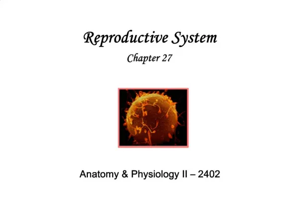
Development of the Human Embryo • Human Ovum • Corona Radiata • Zona pellucida • Egg-plasma with yolk • Female pronucleus • Spermatozoid
Development of the Human Embryo • Entering of the Spermatozoid • Zona pellucida • Fertilization membrane (Between 1 and 2 the perivitelline space). • Protoplasm • Female pronucleus
Development of the Human Embryo • Formation of the Second Polocyte • Chromosomes • Spindle • Sperm nucleus • First polar body
Development of the Human Embryo • Nuclear Copulation • Female pronucleus with female chromosomes • Sperm nucleus with male chromosomes • Centrosome • First and second polar body
Development of the Human Embryo • Female Pronucleus moves towards the Center • Female pronucleus • Sperm nucleus • First and second polar body
Development of the Human Embryo Unicellular Stage 1. First and second polar body.
Development of the Human Embryo Bicellular stage 1. First and second polar body
Development of the Human Embryo Four-cell stage 1. First and second polar body.