Download
1 / 50
580 likes | 934 Views
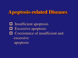
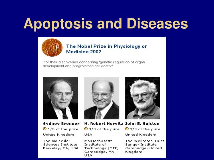
Apoptosis and Diseases. Concept Apoptotic process and changes Key molecules and Major pathways Techniques to detect apoptosis Apoptosis-related diseases Insufficient apoptosis in diseases Excessive apoptosis in diseases Coexistence of insufficient and excessive apoptosis in diseases

E N D
Concept • Apoptotic process and changes • Key molecules and Major pathways • Techniques to detect apoptosis • Apoptosis-related diseases • Insufficient apoptosis in diseases • Excessive apoptosis in diseases • Coexistence of insufficient and excessive apoptosis in diseases • Principles of treatment
What is Apoptosis? • Apoptosis refers to the process in which the dying procedures that have been in advance deposited in cell are triggered by various causes from in vitro and in vivo, and eventually cause cell death. • Programmed cell death(PCD)
Causes process of Apoptosis Stimulatory Factors Inhibitory Factors Physiological: FasL; Pathological: glutamate, free radicals; therapeutics. Physiological: GFs, estrogen, etc; Pathological: virus; chemicals, etc. Initiation Regulation Execution Conserved Phagocytosis
Apoptotic changes---Morphological changes in apoptosis ---Biochemical Changes in Apoptosis
Morphological changes in apoptosis Cell membrane Cytoplasm Cell nucleus Apoptotic body Phagocytose condensation margination Normal Cell Apoptotic Cell Budding Apoptotic Bodies Changes of Cell membrane
Biochemical Changes in Apoptosis • Caspase activation • Endonuclease activation
Caspases: cysteine-containing aspartate-specific proteases • Most apoptotic proteolytic cleavage results from the action of caspases • Caspases are activated by proteolytic cleavage • Removal of prodomain and linker region • Assembly of the large and small subunits into an active enzyme complex • Two heterodimers interacting via the small subunits to form a tetramer with two catalytic sites • Family members>14
Caspase-deficient mice Knockout Phenotype Caspase-1 Viable; impaired processing of IL-1; resistant to endotoxic shock. Caspase-2 Viable; excess numbers of female germ cells; oocytes resistant to chemotherapeutic drugs; B lymphoblasts resistant to granzyme B; accelerated death of facial neurons during development and of sympathetic neurons deprived of NGF. Caspase-3 Lethality at 3–5 weeks of age; defective neuronal apoptosis; T cells resistant to antigen-induced death; abnormal apoptotic morphology in dying cells. Caspase-8 Lethality around E12.5; hyperemia and abnormal heart muscle development; MEFs resistant to TNF, Fas and DR3 but sensitive to UV irradiation, etoposide, staurosporine, serum deprivation. Caspase-9 Perinatal lethal; impaired neuronal apoptosis; ES cells, MEFs and thymocytes generally resistant to intrinsic death stimuli such as DNA damage, though resistance depends on cell type. Caspase-11 Viable; impaired processing of caspase-1, IL-1; resistant to endotoxic shock. Caspase-12 Viable; embryonic fibroblasts are resistant to ER stress.
Caspases activation • Death receptor pathway: caspase 8 • Mitochondrial pathway: caspase 9 • ER stress pathway: caspase 12 (CAD: caspase-activated deoxyribonulease)
Apoptotic substrates ( DNA-PKCS, DNA protein kinase catalytic subunit; HnRNP, heteronuclear ribonucleoproteins; ICAD,inhibitor caspase activated deoxyribonuclease; FAK,focal adhesion kinase; GAS,growth arrest specific gene-2; GDI, GDP dissociation inhibitor; NuMA,nuclear mitotic apparatus; PAK,p21 activated kinase;PARP, poly(ADP-ribose) polymerase; cPLA2, cytoplasmic phospholipase A2; RFC-140, replication factor C; SAF-A,scaffold attachment factor-A; U1-70kDa, U1-specific 70-kDa protein; )
Role of Endonuclease:degrade DNA H1 Signaling activation Endonuclease Ca2+ Mg2+ Zn2+ 180-200 bp
Regulators of ApoptosisBcl2 family proteinsIAP proteins (Inhibitors of Apoptosis)
Bcl2 family: killers and protectors Two groups (>15 members) ---Suppressors of apoptosis: Bcl2, BclXL, BclW, Bag1, Mcl1, A1, etc ---Activators of apoptosis: Bax, Bok, Hrk, Bnip3, Bim, Bik, BclXs, Bik, Blk, Bid, Bak, Bad, etc. Forms heterodimers to keep the balance between apoptosis and survival • On the cytoplasmic face of the outer mitochondrial membrane, endoplasmic reticulum, and nuclear envelope • In hematopoietic cell, epithelial cell, lymphocyte, nerve cell, and various cancer cells
Bcl-2:regulate the release of pro-apoptotic molecules from mitochondriaStructure of Bcl-2 family TM TM: transmembrane region; BH: Bcl-2 homology
Bax: • Apoptotic stimuli induce translocation of Bax from cytosol to mitochondria • Bax seems to create pores in the outer membrane of mitochondria of sufficient size to allow cytochrome C to escape
Inhibitors of Apoptosis-------IAPs family members: c-IAP1,c-IAP2,XIAP,NAIP,survivin;----preventing some procaspases activation, or inhibiting caspase activity.
Apoptosis pathways and related genes • Death Receptor induced apoptosis • Mitochondria – Integrator of Apoptosis • others
Death Factor Family and Death Receptor Family
Fas ( factor associated suicide): • Homologous cytoplasmic domain: death domain (DD) • Interacts with each other through DD • Anti-apoptotic pathway: NF-kB pathway • TNF rarely induces apoptosis unless protein synthesis is inhibited • Decoy receptors Death receptor induced apoptosis
Apoptosis signaling by CD95 (Left) , TNFR1(Middle), and DR3(Right)
Apoptosis signaling by DR4 and DR5 and its modulation by decoy receptors
Three Types of Killing by the Fas and FasL System • Activation-induced suicide of T cells • CTL-mediated killing of target cells • Killing of inflammatory cells in immune privilege sites and killing of CTL by tumor cells
p53-Inducible Apoptosis Related Genes • Scotin: localized to the ER and the nuclear membrane • PERP:similarity to PMP-22/gas3 tetraspan membrane protein • NOXA: A member of Bcl-2 family • BAX • KILLERS/DR5 • FAS • P53AIP1: p53-regulated apoptosis-inducing protein 1, leads to apoptosis via dissipation of mitochondrial△ψm • PIDD: A new death domain containing protein • PIG: P53 induced genes,related to ROS production • IGFBP
Current models of the intracellular pathways leading to trophic factor mediated cell survival in mammalian cells
Current models of the intracellular pathways leading to apoptosis induced by withdrawal of trophic factor
Four patterns of death: from apoptosis to necrosis Apoptosis is observed almost exclusively when caspases, in particular caspase-3, are activated. Apoptosis-like PCD chromatin condensation less compact without other apoptotic features “caspase-independent apoptosis” Necrosis-like PCD no chromatin condensation with chromatin clustering to speckles. Usually involves specialized caspase-independent signalling pathways. “aborted apoptosis” Accidental necrosis/cell lysis associated with cellular oedema (organelle swelling) and devoid of zeiosis
Techniques to detect apoptosis Morphological studies DNA ladder TUNEL Flow cytometry Externalization of Phosphatidylserine Activation of caspases and cleavage of their substrates
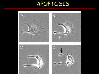
Ultrastructural feathers of Normal and Apoptotic Cell Induced Apoptosis of Cultured Rat Hepatocytes
DNA Ladder Pattern Seen in Diospyrin diethyl ether Induced Apoptotic Cell
Fragmented DNA can be labeled byTerminal deoxynucleotidyl transferase (TdT) mediated deoxyUridine Nucleotide (dUTP)-End Labeling (TUNEL)
Phosphatidylserine on the surface of apoptotic cells[stained with annexin V (green)] Due to: Caspase-3-mediated cleavage and activation of scramblase PKC activation Inactivated amino- phospholipid translocase
Activation of Caspase 3 and Cleavage of Its Substrates, PARP and D4-GDI