Download
1 / 10
120 likes | 295 Views
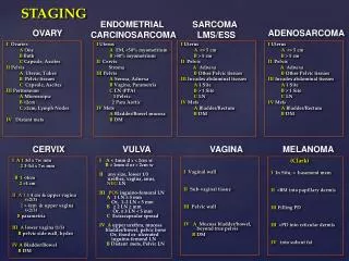
Staging OF BRONCHOGENIC CA. NSCLC STAGING. TNM CLASSFICATION. IA - T1N0M0 IB - T2N0M0 IIA - T1N1M0 IIB - T2N1M0 or T3N0M0 IIIA - T1-3N2M0 or T3N1M0 IIIB - Any T4 or any N3M0 IV - Any M1. Adenocarcinoma Squamous cell carcinoma Large cell carcinoma T – Primary tumor

E N D
NSCLC STAGING TNM CLASSFICATION IA - T1N0M0 IB - T2N0M0 IIA - T1N1M0 IIB - T2N1M0 or T3N0M0 IIIA - T1-3N2M0 or T3N1M0 IIIB - Any T4 or any N3M0 IV - Any M1 • Adenocarcinoma • Squamous cell carcinoma • Large cell carcinoma • T – Primary tumor • N – Regional Lymph nodes • M - Metastasis
NSCLC STAGINGTNM CLASSIFICATION T – PRIMARY TUMOR METHODS OF STAGING CXR – demonstrates peripheral lesions pleural effusion, direct extension to the ribs, phrenic nerve involvement with elevation of a hemidiaphragm, or mediastinal widening due to lymphadenopathy CT SCAN – contrast enhanced thorax and abdomen that includes the liver and adrenal glands Lung window settings: the maximum long axis largest diameter perpendicular to the long axis
NSCLC STAGINGTNM CLASSIFICATION T – PRIMARY TUMOR METHODS OF STAGING MRI superior to CT in assessing the pericardium, heart, and great vessels coronal images are useful in demonstrating the extent of tumor in the subcarinal region, aortopulmonary window & SVC PET SCAN superior to CT in differentiating between malignant and benign tumors
NSCLC STAGINGTNM CLASSIFICATION N – REGIONAL LYMPH NODES METHODS OF STAGING CXR inferior to CT in the detection of mediastinal lymph node metastases, SN of 10-30% only CT SCAN spiral or multisection CT, thin (5-mm) sections SN and SP of 40-84% and 52-80% respectively MRI comparable to those used at CT distinguish nodes from vessels without IV contrast enhancement PET SCAN is superior to CT
NSCLC STAGINGTNM CLASSIFICATION M - METASTASIS METHODS OF STAGING CT SCAN Liver, adrenal, brain and lung metastases Technetium-99m (99mTc) radionuclide bone scanning Bone metastases PET SCAN Adrenal metastases
SCLC Staging LIMITED STAGE DISEASE EXTENSIVE STAGE DISEASE disseminated nature of SCLC makes whole-body survey techniques suitable for its evaluation 99mTc-labeled monoclonal antibody fragment NR-LU-10 detect an antigen present in most small cell cancers whole-body FDG-PET Detects nodal disease. combined MRI of the brain, spine, abdomen, and pelvis enables comprehensive staging with a single modality • confined to 1 hemithorax • ( includes ipsilateral, contralateral, and/or supraclavicular nodes) • CXR; CT of the thorax, liver, and adrenal glands; cranial CT; bone scintigraphy, bone marrow aspiration