Download
1 / 1
10 likes | 127 Views
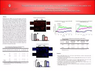
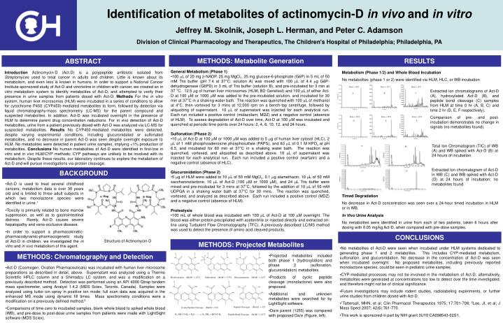
Structure of Actinomycin-D. Hydroxylation (M+H) + = 1271. Sulfonation (M+H) + = 1351. Glucuronidation (M+H) + = 1457. Peptide Cleavages (M+H) + = 327. Cyclic Peptide Cleavage (M+H) + = 971. Peptide Bond Cleavage (M+H) + = 1273.

E N D
Structure of Actinomycin-D Hydroxylation (M+H)+ = 1271 Sulfonation (M+H)+ = 1351 Glucuronidation (M+H)+ = 1457 Peptide Cleavages (M+H)+ = 327 Cyclic Peptide Cleavage (M+H)+ = 971 Peptide Bond Cleavage (M+H)+ = 1273 R1-NH-CO-R2 + H2O R1-NH2 + HOOC-R2 Identification of metabolites of actinomycin-D in vivo and in vitro Jeffrey M. Skolnik, Joseph L. Herman, and Peter C. Adamson Division of Clinical Pharmacology and Therapeutics, The Children's Hospital of Philadelphia; Philadelphia, PA Da/e = 1255 Da/e = 1271 Da/e = 1273 A B C Da/e = 1255 Da/e = 1271 Da/e = 1273 D E F METHODS: Metabolite Generation RESULTS ABSTRACT • General Metabolism (Phase 1) • 100 L of 20 mg -NADP, 25 mg MgCl2, 35 mg glucose-6-phosphate (G6P) in 5 mL of 50 mM Tris buffer (pH 7.4 at 37°C; solution A) was mixed with 100 L of 4.4 g G6P-dehydrogenase (G6PD) in 3 mL of Tris buffer (solution B), and pre-incubated for 3 min at 37 °C. 12.5 g of human liver microsomes (HLM; BD Genetest) and 100 L of either Act-D at 100 M or 1000 M was added to the pre-incubated solution, and incubated for 30 min at 37°C in a shaking water bath. The reaction was quenched with 100 L of methanol at 4°C, then vortexed for 3 mins at 12,000 rpm on a bench-top centrifuge, followed by aliquotting of supernatant. 10 L of supernatant was injected for each analytical run. Each run included a positive control (midazolam; MDZ) and a negative control (absence of HLM). To assess degradation of Act-D over time, Act-D at 100 M was incubated and quenched at periodic time points over 24 hours: 2, 4, 6, 10, 12, and 24 hours. • Sulfonation (Phase 2) • 10 L of Act-D at 100 M or 1000 M was added to 5 g of human liver cytosol (HLC), 2 L of 1 mM phosphoadenosine phosphosulfate (PAPS), and 83 L of 0.1 M KPO4 at pH 6.5, and incubated for 60 min at 37°C in a shaking water bath. The reaction was quenched, vortexed, and aliquotted as described above. 10 L of supernatant was injected for each analytical run. Each run included a positive control (warfarin) and a negative control (absence of HLC). • Glucuronidation (Phase 2) • 5 g of HLM were added to 10 L of 50 mM MgCl2, 0.1 g alamethacin, 10 L of 50 mM saccharonolactone, 10 L of Act-D (100 M or 1000 M), and 24 L Tris buffer were mixed and pre-incubated for 3 mins at 37°C, followed by the addition of 10 L of 50 mM UDPGA in a shaking water bath at 37°C for 30 mins. The reaction was quenched, vortexed, and analyzed as described above. Each run included a positive control (MDZ) and a negative control (absence of HLM). • Proteolysis • 100 mL of whole blood was incubated with 100 L of Act-D at 100 M overnight. The blood was either protein precipitated with acetonitrile or injected directly and extracted on-line using Turbulent Flow Chromatography (TFC). A previously described LC/MS method was used to detect the presence of amino acid cleaved products. Introduction Actinomycin-D (Act-D) is a polypeptide antibiotic isolated from Streptomyces used to treat cancer in adults and children. Little is known about its metabolism, and even less is known in humans. In order to support a National Cancer Institute-sponsored study of Act-D and vincristine in children with cancer, we created an in vitro metabolism system to identify metabolites of Act-D, and attempted to verify their presence in urine samples from patients dosed with Act-D. Methods For the in vitro system, human liver microsomes (HLM) were incubated in a series of conditions to allow for cytochrome P450 (CYP450)-mediated metabolites to form, followed by detection via liquid chromatography-mass spectrometry (LC-MS) for the parent compound and suspected metabolites. In addition, Act-D was incubated overnight in the presence of HLM to determine parent drug concentration reductions. For in vivo detection of Act-D metabolites, urine from a patient previously dosed with Act-D was analyzed via LC-MS for suspected metabolites. Results No CYP450-mediated metabolites were detected, despite varying experimental conditions, including glucuronidated or sulfonated metabolites, and no decrease in parent Act-D was seen despite overnight exposure to HLM. No metabolites were detected in patient urine samples, implying <1% production of metabolites. Conclusions No human metabolites of Act-D were identified in first-line in vitro and in vivo HLM/CYP methods; CYP pathways are unlikely to be involved with its metabolism. Despite these results, our laboratory continues to explore the metabolism of Act-D and will pursue investigations via protein cleavage. Metabolism (Phase 1/2) and Whole Blood Incubation No metabolites (phase 1 or 2) were identified via HLM, HLC, or WB incubation Extracted ion chromatograms of Act-D (A), hydroxylated Act-D (B), and peptide bond cleavage (C) samples from HLM at time 0 hr (A, B, C) and time 2 hr (D, E, F respectively). Comparison of pre- and post- incubation demonstrates no change in signals (no metabolites found). Total Ion Chromatogram (TIC) of WB (A) and WB spiked with Act-D (B) at 24 hours of incubation. Extracted Ion chromatogram of Act-D in WB (C) and WB spiked with Act-D (D) at 24 hours of incubation; no metabolites found. BACKGROUND • Act-D is used to treat several childhood cancers; metabolism data is over 30 years old and is limited to three adult subjects in which two monolactone species were identified in urine.1 • Toxicity is primarily related to bone marrow suppression, as well as to gastrointestinal distress. Rarely, Act-D causes severe hepatopathy and veno-occlusive disease. • In order to support a pharmacokinetic-pharmacodynamic-pharmacogenetic study of Act-D in children, we investigated the in vitro and in vivo metabolism of this agent. Timed Degradation No decrease in Act-D concentration was seen over a 24-hour timed incubation in HLM or in WB. In Vivo Urine Analysis No metabolites were identified in urine from each of two patients, taken 6 hours after dosing with 0.05 mg/kg Act-D, when compared with pre-dose samples. CONCLUSIONS METHODS: Projected Metabolites • No metabolites of Act-D were seen when incubated under HLM systems dedicated to generating phase 1 and 2 metabolites. This includes CYP-mediated metabolism, sulfonation, and glucuronidation. No decrease in the concentration of Act-D was seen when incubated overnight. No proposed metabolites, including previously reported monolactone species, could be seen in pediatric urine samples. • CYP-mediated processes may not be involved in the metabolism of Act-D; alternatively, metabolites were generated at concentrations too low to detect over the time investigated, and therefore might not be of clinical significance. • Future investigations may include rodent studies, radiolabeling experiments, or further urine studies from children dosed with Act-D. • 1Tattersall, MHN, et al; Clin Pharmacol Therapeutics 1975; 17:701-708; 2Lee, JI, et al; J Mass Spect 2007; 42(6):761-770. • This work is sponsored in part by NIH grant 3U10 CA098543-02S1. METHODS: Chromatography and Detection • Projected metabolites included both phase 1 (hydroxylation) and phase 2 (sulfonation, glucuronidation) metabolites. • Products of cyclic peptide cleavage (monolactone) were also proposed. • Additional and unknown metabolites were searched for by LightSight software. • Da/e parent (1255) was compared with proposed Da/e (Figure, left). • Act-D (Cosmegen; Ovation Pharmaceuticals) was incubated with human liver microsome preparations as described in detail, above. Supernatant was analyzed using a Thermo Scientific HPLC column and a Shimadzu LC system, and was a modification on a previously described method. Detection was performed using an API 4000 Qtrap tandem mass spectrometer, using Analyst 1.4.2 (MDS Sciex, Toronto, Canada). Samples were analyzed using turbo ion spray in positive ion mode; full scan data was acquired in the enhanced MS mode using dynamic fill times. Mass spectrometry conditions were a modification on a previously defined method.2 • Comparisons of time-zero to incubated samples, blank whole blood to spiked whole blood (WB), and pre-dose to post-dose urine samples from patients were made with LightSight software (MDS Sciex).