Download
1 / 10

E N D
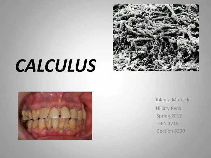
CALCULUS Jolanta Moscicki Hillary Pena Spring 2012 DEN 1218 Section 8230
Calculus is a bacterial plaque that has been mineralized.Itis a deposit of calcium phosphate salts. It has variety of appearance on dental radiograph. The bitewing shows subgingival calculus which is visible as cervical spurs upon the proximal tooth surface in the posterior segments, on the maxillary and mandible first and second molars.
Calculus may appear as a ring-like radiopacity around the root of mandibular teeth
Diabetes mellitus can cause dysfunction of the secretory ability of the salivary gland. Diabetic patient have an increased salivary calcium phosphates and protein concentration compared to non-diabetic. Here we can see large mass of dental calculus that is covering the surface of two remaining teeth.
Calculus may appear nodular, as seen here on the distal side of the third molar, beneath the crown margin.
Salivary calculus ( Sialolithiasis) is a salivary duct obstruction. It can be caused by trauma or local inflammation, chronic disease ,infection. Dental radiograph of the sublingual space reveals the size and location of the salivary calculus
Subgingival calculus appears as irregular radiopaque projections on the maxillary incisors.
Ring-like calculus (radiopaque line) on mandibular canine and first premolar.
References: Dental Radiography by J.M. Iannucci (2006) www.google.com/ images from hillam .net www.rickwilsondmd.typepad.com www. endodonticspecialist.blogs.com www.rootcanaltreatment.blogspot.com www.nature.com/bdj/journal/v205/n11 www.google .com/images from health-7.com