Download
1 / 47
520 likes | 778 Views
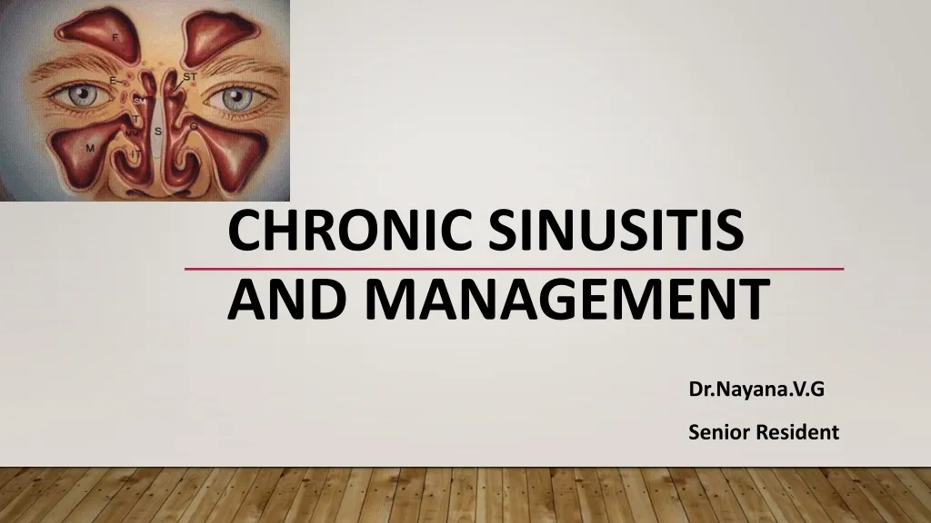
Chronic sinusitis and management. D r .n ayana .v.g S enior r esident. Specific learning objective. 1. To learn the definition, etiology, pathophysiology symptoms and signs of chronic rhinosinusitis 2.To learn the diagnostic methods medical and surgical management of chronic rhinosinusitis

E N D
Chronic sinusitis and management Dr.nayana.v.g Senior resident
Specific learning objective 1. To learn the definition, etiology, pathophysiology symptoms and signs of chronic rhinosinusitis 2.To learn the diagnostic methods medical and surgical management of chronic rhinosinusitis 3.To know the surgical steps of FESS
Definition: Rhinosinusitis Task Force • Inflammation of the nose & PNS characterized by two or more symptoms one of which should be either nasal blockage or nasal discharge • +/- facial pain/pressure • +/-reduction or loss of smell • DNE :Polyps or mucopurulent discharge or edema primarily in middle meatus • +/-CT findings of mucosal changes in OMC or sinuses
Chronic sinusitis -Definition • Duration:>12 wks • Persistent inflammatory changes on imaging > 4wks after starting treatment
Etiology • Mechanical (OMC) Obstruction • Infections Micro organisms – Secondary invaders • Allergy • Decreased mucocilliary function • Dental disease
Micro organisms • Bacteria – Staphylococcus Streptococcus pseudomonas M.catarrhalis Gram negative • Fungi
Major & Minor signs and symptoms in diagnosis of chronic rhinosinusitis Major Factors • Nasal obstruction/blockage • Nasal discharge/purulence • Facial pain/pressure • Hyposmia/ anosmia Minor Factors: • Headache • Fever • Fatigue • Halitosis • Dental pain • Cough • Ear pain/ pressure/fullness
Clinical Diagnosis of Rhinosinusitis 2 or more major factors 1 major & 2 minor factors
Sinus pain or pressure • Frontal : Frontal Headache • Maxillary : Mid-face, maxillary tooth pain • Ethmoid : Retro-orbital pain • Sphenoid : Nonspecific pain with radiation • Pain increases on bending forward, in late morning, with mastication
Management - Diagnosis and Treatment • Routine physical examination of nose and sinus • Diagnostic Nasal endoscopy • Sinus aspiration and culture
Physical examination of nose and sinus • Edema and erythema of nasal mucosa • Turbinate Hypertrophy • Purulent discharge from lateral wall • Sinus tenderness.
Sinus aspiration and culture • Correlation of routine nasal culture and sinus culture are poor • Endoscopically guided aspiration from medial meatus do correlate with sinus culture
RADIOLOGY Indications • Chronic, refractory or recurrent sinusitis • High risk patients (DM, Immunosuppressed) • Complicated sinusitis Studies • X Ray • CT (Gold standard)
Findings suggestive of Sinusitis • Opacification • Air fluid levels Efficacy in Acute Sinusitis Sensitivity 76% Specificity 79% Not effective in children under 3
Sinus CT Advantages More sensitive than X Ray Better anatomy definition Sphenoid, Ethmoid and OMC Preparation for Surgery
CT scan • 3-4 weeks after medical therapy. • 2-5 mm coronal & axial cuts. • Assess sino nasal anatomy & disease pattern • Anatomic variations(Meyers & Valvassori) • Roadmap for surgical intervention
Medical manangement of Chronic Sinusitis AIM OF TREATMENT • To reduce symptoms and signs, improve patients quality of life and prevent disease progression and/or recurrence.
Medical management of chronic rhinosinusitis 1. Avoidance of allergens2.Avoidance of irritants 3. Antibiotics 4.Antihistamines 5. Decongestants 6. Corticosteroids 7.Sinus irrigation
Treatment to improve OMC drianage • Decongestant • Nasal Douching • Topical corticosteroids • Review at 6 week,if no improvement -> CT scan
Surgery for maxillary sinusitis • Antral puncture • Intranasal antrostomy • Caldwell luc surgery • FESS
Surgeries for Chronic Frontal Sinusitis • Frontal sinus trephination • External frontoethmoidectomy • Osteoplastic flap operation • FESS
Surgeries for Ethmoid Sinusitis • Intranasal ethmoidectomy • External ethmoidectomy
FUNCTIONAL ENDOSCOIC SINUS SURGERY • The concept is to surgically remove inflamed tissue and involved bone from critical points in the mucociliary clearance pathways
AIMS of FESS • Restore paranasal sinus function. • Re-establish the physiologic pattern of mucociliary clearance & ventilation. • Remove diseased mucosa & bone. • Preserve normal tissue . • Judiciously widen the natural ostia.
Anaesthesia Local Anaesthesia • Awake patients can report manipulation of the orbital periosteum or dura. • Young patients undergoing primary sinus surgery that lasts less than 2 hours General anaesthesia • Anxious patient • Operation in a child • Anticipation of a long procedure.
Preparation & positioning • Supine position • Local anaesthetic- 2% lignocaine + adrenaline (1:100,000) • Nasal septum, inferior turbinate, and middle turbinate are injected. • Cotton pledgets soaked in conc adrenaline kept in nasal cavity. • Throat pack . • Eyes lubricated & taped in GA
Nasal endoscopy • Look for landmarks • Condition of the mucosa, • The presence of any polyps or pus • Structural abnormalities identified • Additional injections of local anesthetic can be given
uncinectomy A ball-tipped probe is slid into the infundibulum Identified with the 0-degree endoscope Middle turbinate can be gently rotated using a Freerelevator.
A backbiter punch is used to incise the uncinate process. Between the inferior one third and superior two thirds of the uncinate process Incision is continued anteriorly until the harder lacrimal bone is encountered
Inferior aspect is rotated medially with a probe and is removed with a Microdebrider
Maxillary Antrostomy • A Lusk probe used to identify the ostium • Ostium widened posteriorly & inferiorly. • Probe should not be forced into the ostium. • Accessory ostium and natural ostium joined
Anterior Ethmoidectomy • Ethmoid bulla is the largest cell. • Anterior and medial walls then taken down. • Lateral wall of the ethmoid bulla is the lamina papyracea. • Mucosa should be preserved laterally • Agger nasi and supra bullar cells opened
Sphenoid Sinusotomy • Ostium is located approximately halfway between the superior and inferior borders of the anterior wall. • Natural ostium can be located by gentle sliding of a probe along the anterior wall. • The probe should never be forced.
Sphenoid Sinusotomy • Ostium , once identified, should be widened. • Forceps inserted, opened & pulled out to out fracture the anterior wall. • Disease removed from the sinus.
Frontal Sinusotomy • If not diseased, it should be left alone. • Minimize mucosal injury and prevent middle turbinate lateralization. • Remnants of the uncinate process should be carefully removed. • Mucosal stripping in the region of frontal recess causes recurrence of disease.
Small bone fragments removed. • Hemostasis attained • The areas of the sphenopalatine, posterior septal, anterior ethmoidal, and posterior ethmoidal arteries are carefully examined. • Pack can be kept in the middle meatus to prevent lateralisation.
Summary • Rhinosinusitis persisting more than 12 weeks • Nasal obstruction, discharge, Facial pain and hyposmia are the major symptoms • CT Nose and PNS is the gold standard investigation • Initially treated with medical management if it fails endoscopic sinus surgery is the treatment of choice
Reference.. 1.Disease of Ear, Nose, Throat &Head and Neck surgery – PL Dhingra, Shruthi Dhingra Acknowledgement Images from google images and text book(Disease of Ear, Nose, Throat &Head and Neck surgery – PL Dhingra, Shruthi Dhingra)