Download
1 / 32
320 likes | 720 Views
Russell_2e_IRCD_Chapter_2. 2. Review Principal Points. What is the sugar for DNA? RNA?What are the possible bases?What are the possibilities for genetic arrangement?How are bacterial chromosomes compacted?Define karyotypeWhat is the composition of eukaryotic genomes; what is constant between ce

E N D
1. Russell_2e_IRCD_Chapter_2 1 Chapter 2: DNA The Genetic Material Linnea Fletcher Ph.D.
BIOL 2316
2. Russell_2e_IRCD_Chapter_2 2 Review Principal Points What is the sugar for DNA? RNA?
What are the possible bases?
What are the possibilities for genetic arrangement?
How are bacterial chromosomes compacted?
Define karyotype
What is the composition of eukaryotic genomes; what is constant between cells of a particular organism and what varies?
What is the role of the centromere?
What is the composition of the telomere?
Compare and contrast the prokaryotic genome with the eukaryotic genome.
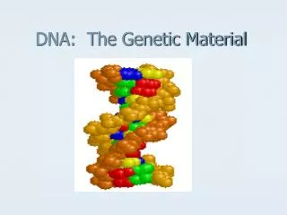
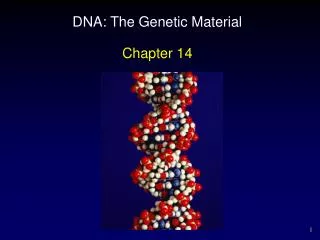
3. Russell_2e_IRCD_Chapter_2 3 DNA double helix. Left, end view; right, side view. The red balls represent oxygen; the yellow phosphorous; the purple carbon; and the blue nitrogen.DNA double helix. Left, end view; right, side view. The red balls represent oxygen; the yellow phosphorous; the purple carbon; and the blue nitrogen.
4. Russell_2e_IRCD_Chapter_2 4 The Search for Genetic Material What are the three principal characteristics for genetic materials?
Stable form
Replicate accurately
Capable of change
5. Russell_2e_IRCD_Chapter_2 5 Understand the experiments performed by:
1928 Griffith
1944 Avery, McCarty, & MacLeod
1947 Chargaff
1954 Wilkins, Franklin, Watson, & Crick
6. Russell_2e_IRCD_Chapter_2 6 Figure 2.1 Electron micrograph of the bacterium Streptococcus pneumoniae. Figure 2.1 Electron micrograph of the bacterium Streptococcus pneumoniae.
7. Russell_2e_IRCD_Chapter_2 7 Figure 2.2 Griffith�s transformation experiment. Mice injected with type IIIS pneumococcus died, whereas mice injected with either type IIR or heat-killed type IIIS bacteria survived. When injected with a mixture of living type IIR and heat-killed type IIIS bacteria, however, the mice died. Figure 2.2 Griffith�s transformation experiment. Mice injected with type IIIS pneumococcus died, whereas mice injected with either type IIR or heat-killed type IIIS bacteria survived. When injected with a mixture of living type IIR and heat-killed type IIIS bacteria, however, the mice died.
8. Russell_2e_IRCD_Chapter_2 8 Figure 2.3 Experiment which showed that DNA, not RNA, was the transforming principle. When the nucleic acid mixture of DNA and RNA was treated with ribonuclease (RNase), S transformants still resulted. However, when the DNA and RNA mixture was treated with deoxyribonuclease (DNase), no S transformants resulted. (Note: R colonies resulting from untransformed cells were present on both plates; they have been omitted from the drawings.) Figure 2.3 Experiment which showed that DNA, not RNA, was the transforming principle. When the nucleic acid mixture of DNA and RNA was treated with ribonuclease (RNase), S transformants still resulted. However, when the DNA and RNA mixture was treated with deoxyribonuclease (DNase), no S transformants resulted. (Note: R colonies resulting from untransformed cells were present on both plates; they have been omitted from the drawings.)
9. Russell_2e_IRCD_Chapter_2 9
10. Russell_2e_IRCD_Chapter_2 10 Figure 2.12 X-ray diffraction analysis of DNA. (a) Rosalind Franklin and Maurice H. F. Wilkins (photographed in 1962, the year he received the Nobel Prize shared with Watson and Crick). (b) The X-ray diffraction pattern of DNA that Watson and Crick used in developing their double-helix model. The dark areas that form an X shape in the center of the photograph indicate the helical nature of DNA. The dark crescents at the top and bottom of the photograph indicate the 0.34-nm distance between the base pairs.Figure 2.12 X-ray diffraction analysis of DNA. (a) Rosalind Franklin and Maurice H. F. Wilkins (photographed in 1962, the year he received the Nobel Prize shared with Watson and Crick). (b) The X-ray diffraction pattern of DNA that Watson and Crick used in developing their double-helix model. The dark areas that form an X shape in the center of the photograph indicate the helical nature of DNA. The dark crescents at the top and bottom of the photograph indicate the 0.34-nm distance between the base pairs.
11. Russell_2e_IRCD_Chapter_2 11 What did the X-ray diffraction patterns analyzed by Rosalind Franklin INITIALLY reveal about the DNA molecule?
It is of uniform diameter about 2 nm wide and has a highly repetitive structure that repeats every 3.5 nm
It is a helical molecule with paired bases in the center
It is double-stranded with antiparallel strands
It is acidic, phosphate-rich, and very large
It contains the hereditary information
12. Russell_2e_IRCD_Chapter_2 12 Figure 2.11 James Watson (left) and Francis Crick (right) in 1993 at a 40th-anniversary celebration of their discovery of the structure of DNA and in 1953 with the model of DNA structure. Figure 2.11 James Watson (left) and Francis Crick (right) in 1993 at a 40th-anniversary celebration of their discovery of the structure of DNA and in 1953 with the model of DNA structure.
13. Russell_2e_IRCD_Chapter_2 13 . An understanding of the molecular basis of inheritance allows us to develop a detailed knowledge of how this works.
Describe the structural features of DNA and RNA. How are they similar, and how are they different?
How do purines differ from pyrimidines? Which base-pair with each other?
How do nucleosides differ from nucleotides?
What type of covalent bonding combines the monomers into polymers?
Why is the DNA double helix referred to as being �antiparallel�?
14. Russell_2e_IRCD_Chapter_2 14
15. Russell_2e_IRCD_Chapter_2 15 Figure 2.8 Structures of deoxyribose and ribose, the pentose sugars of DNA and RNA, respectively. The difference between the two sugars is highlighted. Figure 2.8 Structures of deoxyribose and ribose, the pentose sugars of DNA and RNA, respectively. The difference between the two sugars is highlighted.
16. Russell_2e_IRCD_Chapter_2 16 Figure 2.9 Structures of the nitrogenous bases in DNA and RNA. The parent compounds are purine (top left) and pyrimidine (bottom left). Differences between the bases are highlighted. Figure 2.9 Structures of the nitrogenous bases in DNA and RNA. The parent compounds are purine (top left) and pyrimidine (bottom left). Differences between the bases are highlighted.
17. Russell_2e_IRCD_Chapter_2 17 Figure 2.10 Chemical structures of DNA and RNA. (a) Basic structures of DNA and RNA nucleosides (sugar plus base) and nucleotides (sugar, plus base, plus phosphate group), the fundamental building blocks of DNA and RNA molecules. Here, the phosphate groups are orange, the sugars are red, and the bases are brown. (b) A segment of a polynucleotide chain, in this case a single strand of DNA. The deoxyribose sugars are linked by phosphodiester bonds (shaded) between the3� carbon of one sugar and the 5� carbon of the next sugar.Figure 2.10 Chemical structures of DNA and RNA. (a) Basic structures of DNA and RNA nucleosides (sugar plus base) and nucleotides (sugar, plus base, plus phosphate group), the fundamental building blocks of DNA and RNA molecules. Here, the phosphate groups are orange, the sugars are red, and the bases are brown. (b) A segment of a polynucleotide chain, in this case a single strand of DNA. The deoxyribose sugars are linked by phosphodiester bonds (shaded) between the3� carbon of one sugar and the 5� carbon of the next sugar.
18. Russell_2e_IRCD_Chapter_2 18 Figure 2.13 Molecular structure of DNA. (a) Three-dimensional molecular model of DNA as prepared by Watson and Crick. (b) Stylized representation of the DNA double helix. (c) Chemical structure of a segment of double-stranded DNA. Figure 2.13 Molecular structure of DNA. (a) Three-dimensional molecular model of DNA as prepared by Watson and Crick. (b) Stylized representation of the DNA double helix. (c) Chemical structure of a segment of double-stranded DNA.
19. Russell_2e_IRCD_Chapter_2 19 Figure 2.14 Structures of the complementary base pairs found in DNA. In both cases, a purine pairs with a pyrimidine: (a) The adenine�thymine bases, which pair through two hydrogen bonds. (b) The guanine�cytosine bases, which pair through three hydrogen bonds. Figure 2.14 Structures of the complementary base pairs found in DNA. In both cases, a purine pairs with a pyrimidine: (a) The adenine�thymine bases, which pair through two hydrogen bonds. (b) The guanine�cytosine bases, which pair through three hydrogen bonds.
20. Russell_2e_IRCD_Chapter_2 20 Describe the three oligomers that the DNA double helix can assume: A-from, B-form and Z-form. Which has the most compact structure, and which is the most elongated? Which are found in living cells?
21. Russell_2e_IRCD_Chapter_2 21 Figure 2.15 Space-filling models of different forms of DNA. (a) A-DNA. (b) B-DNA. (c) Z-DNA. Figure 2.15 Space-filling models of different forms of DNA. (a) A-DNA. (b) B-DNA. (c) Z-DNA.
22. Russell_2e_IRCD_Chapter_2 22
23. Russell_2e_IRCD_Chapter_2 23 The organization of nucleic acids in genomes varies with the level of complexity of the organism
What form is the chromosome of most prokaryotes? (circular)
How is supercoiling created in a circular chromosome?
Compare the size of the haploid genome (C-value) of humans with other organisms.
What is the C value paradox?
24. Russell_2e_IRCD_Chapter_2 24
25. Russell_2e_IRCD_Chapter_2 25
26. Russell_2e_IRCD_Chapter_2 26
27. Russell_2e_IRCD_Chapter_2 27 Compaction of chromosomes is important in eukaryotes, due to the large size of the eukaryotic genome.
What are the two types of molecular components of chromatin?
What are the most abundant proteins in chromatin and what role do they play in the structure of the chromosome?
How long would the diploid set of human chromosomes from one cell be if they were not compacted and where place end-to-end in a straight line? How does this compare to the diameter of the nucleus of a human cell?
Describe the structures of the 10-nm and 30-nm chromatin fibers.
Describe how scaffold=associated regions (SARs) are arranged by non-histone proteins in a condensed chromosome. About how many looped domains does the average human chromosome have? What is the approximate diameter of the metaphase chromosome?
28. Russell_2e_IRCD_Chapter_2 28
29. Russell_2e_IRCD_Chapter_2 29
30. Russell_2e_IRCD_Chapter_2 30 Figure 2.25 The 30-nm chromatin fiber. (a) Electron micrograph. (b) Solenoid model for the packaging of nucleosomes into the 30-nm chromatin fiber. (H1 is not shown.)
Figure 2.25 The 30-nm chromatin fiber. (a) Electron micrograph. (b) Solenoid model for the packaging of nucleosomes into the 30-nm chromatin fiber. (H1 is not shown.)
31. Russell_2e_IRCD_Chapter_2 31
32. Russell_2e_IRCD_Chapter_2 32 What affect does compaction of a chromosome have on its transcriptional activity?
Compare the staining of euchromatic, compared to heterochromatic, regions of a chromosome by dyes.
Which is transcriptionally more active: euchromatin or heterochromatin?
What are two types of constitutive heterochromatin and what roles do they play? What structural features do they share, and how are they different?
What is an example of facultative heterochromatin?
33. Russell_2e_IRCD_Chapter_2 33 To make learning easier: Make a table that compares the copy number and the relative amounts of unique-sequence, moderately repetitive, and highly repetitive DNA in the human genome.
In a separate column, indicate which category has these of DNA: genes, SINEs, LINEs, centromeres, telomeres, rRNAs, tRNAs.
How are tandemly repeated sequences different from interspersed repeat sequences of DNA?
Compare the structures of LINEs with that of SINEs. What is the most abundant of each in the human genome, and about how many times are they repeated in the haploid human genome?