Download
1 / 49

E N D
Renal calculi M.Prasad Naidu Msc Medical Biochemistry, Ph.D. research scholar
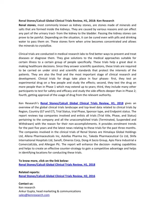
Renal calculi: • The smooth epithileal tissue are formed the hardness by the inorganic and organic substance like kidney--------- stone ( calcium) gall bladder---- stone ( cholesterol oxalates) intestine ------- jejunum (hard substance) Introduction: Urinary calculi are mainly composed of substance normally in urine and may be found in any part of the urinary tract. Their size of an egg. These calculi can be divided into:
Simple calculi • Mixed calculi • Foreign body calculi Formatin: The nucleus for stone can be obtained by the presence of a small lesion. The crystals get deposited on the nucleus and continue to grow. These can some times adhere to the renal papillae. • Substances found in calculi : They are mainly uric acid, urate , triple phosphate, calcium carbonate ,calcium phosphate, calcium oxalates, cholesterol. Cystine calculi have been reported but are extremly rare, and xanthin also form stones ( xanthinuria)
COMPARATIVE INCIDENCES OF FORMS OF URINARY LITHIASIS Stone analysis in Percentage Form of Lithiasis India USA Japan UK Pure Calcium Oxalate 86.1 33 17.4 39.4 Mixed Calcium Oxalate and 4.9 34 50.8 20.2Phosphate Magnesium Ammonium 2.7 15 17.4 15.4Phosphate (Struvite ) Uric Acid 1.2 8.0 4.4 8.0 Cystine 0.4 3.0 1.0 2.8
Inhibitors & Promoters of Stone Formation in Urine INHIBITORS Inhibits crystal Growth - • Citrate – complexes with Ca • Magnesium – complexes with oxalates • Pyrphosphate - complexes with Ca • Zinc Inhibits crystal Aggregation • Glycosaminoglycans • Tamm- Horsfall Protein PROMOTERS • Bacterial Infection • Anatomic Abnormalities – PUJ obst., MSK • Altered Ca and oxalate transport in renal epithelia • Prolonged immobilisation • Increased uric acid levels I.e taking increased purine subs– promotes crystalisation of Ca and oxalate
TYPES OF RENAL / URETER STONES • Common stones: • OXALATE (CALCIUM OXALATE) • PHOSPHATE • URIC ACID / URATE • CYSTINE
Uncommon Stones XANTHINE STONES – (Autosomal Recessive . Def of Xanthine Oxidase leading to Xanthinuria) DIHYDROXY ADENINE STONE – ( Def. of enzyme adenine phospo ribosyl transferase ) SlLICATE STONES – Rare in humans ( excess intake of Antacid with Mg Trisilicate. Mostly in cattle due to ingestion of Sand ) MATRIX - Infection by Proteus - Radiolucent(all calculi have some amt ( 3%) of matrix but matrix calculus has 65% Matrix content in calculi)
Stones – BIO Chemical Constituents • Whewelite – Calcium Oxalate Monohydrate – CaC2O4-H2O • Weddelite - Calcium Oxalate dihydrate – CaC2O4-2H2O • Brushite – Calcium Hydrogen phosphate dihydrate – CaHPO4 2H2O • Whitlockite - TriCalcium Phosphate – Ca2(PO4)2 • Struvite – Magnesium Ammonium hexahydrate – MgNH4PO4-6H2O
D/D of Radiolucent filling defect on IVU in Ureter or Kidney Must Know • Uric Acid Calculus • Matrix Calculus • Sloughed Papilla • Blood Clots • TCC • Renal Cysts • Vascular Lesions Know For Brownie Points • Xanthine Calculus • Hydroxy adenine Calculus • Ephederine Calculus • Infection due to gas forming Org. • Fungal Ball • Tuberculoma • Malacoplakia • Hyper trophied Papilla • Renal pseudo-tumour
OXALATE (CALCIUM OXALATE) • ALSO CALLED MULBERRY STONE • COVERED WITH SHARP PROJECTIONS • SHARP ® MAKES KIDNEY BLEED (HAEMATURIA) • VERY HARD • RADIO - OPAQUE Under microscope looks like Hourglass or Dumbbell shape if monohydrate and Like an Envelope if Dihydrate
Bio chemical test for oxalate stone • Procedure: • Make fine powder • Add 2 to 3 drops of 10% Hcl • Cool it and add pinch Mn O2- do not mix Result: fomation of gas bubbles form bottom
PHOSPHATE STONE • USUALLY ® CALCIUM PHOSPHATE • SOMETIMES ® CALCIUM MAGNESIUM AMMONIUM PHOSPHATE OR TRIPLE PHOSPHATE • SMOOTH ® MINIMUM SYMPTOMS • DIRTY WHITE • RADIO - OPAQUE Calcium Phosphate also called ‘Brushite’ appears like Needle shape under microscope
Bio chemical test for phosphate stone • Procedure: • Make fine powder • Add o.5ml of ammonium molybdate warm over a gas flame Results: formation of yellow precipitate.
PHOSPHATE STONES • IN ALKALINE URINE¯ ENLARGES RAPIDLY¯ TAKE SHAPE OF CALYCES¯ STAGHORN ®
CALCIUM PHOSPHATE STONES • Hyperparathyroidism Ca P • Renal Tubular Acidosis K CO2 • Medullary Sponge Kidney - PTH Hormone Promotes renal production of 1-25-dihyroxycholecalciferol – active Vit.D and also increases absorption of Calcium and decreases Phosphorus absorption from Kidneys
URIC ACID & URATE STONE • HARD & SMOOTH • MULTIPLE • YELLOW OR RED-BROWN • RADIO - LUCENT (USE ULTRASOUND) pKa of uric acid 5.75 at this pH 50% of uric acid insoluble. If pH falls further - uric acid more insoluble
Bio chemical test for urate stone • Murexide test • Procedure: • Make fine powder of the stone by using mortor • Take a pinch of the powder in a test tube • Add 1 drop of 20g/dl Na2 Co3. • Add 2drops of phopho tungstic acid reagent Results : formation of deep blue color. Clinical significance: gout
CYSTINE STONE • AUTOSOMAL RECESIVE DISORDER • USUALLY IN YOUNG GIRLS • DUE TO CYSTINURIA - • CYSTINE NOT ABSORBED BY TUBULES • MULTIPLE • SOFT OR HARD – can form stag-horns • PINK OR YELLOW • RADIO-OPAQUE Under microscope appears like hexagonal or benezene ring – ask for first morning sample
CYSTINE STONE - Management • High Fluid Intake and Alkalanise Urine – dissolve most of the smaller cystine stones • D-Pencillamine or MPG (Mercaptopropionylglycine) binds to cystine that is soluble in urine • Side effects of Pencillamine restricts it use – Allergic rashes, GI problems- Nausea, Vomiting, Diarrhoea • MPG better tolerated • Large obstructive stones – Surgery required first Cyanide Nitroprusside Calorimeteric Test for detecting Cystinuria. If positive do amino acid chromatography
Bio chemical test for cystine stone • Procedure: • Make fine powder • Add 1 drop of ammonium hydrooxide reagent and one drop of Na Cl reagent, wait for 5 min add 2-3 drops of sodium nitroprusside reagent Result: beet red color changes to orange is standing Clinical significance: cystinuria
Cause of Stone Disease • Supersaturation of urine is the key to stone formation • Intermittent supersaturation - Dehydration • Crystal aggregation • Anatomic Abnormailities – PUJ , MSK • Bacterial Infection • Defects in transport of Calcium and Oxalate by Renal epithelia E.Coli infection increases matrix content in urine . Proteus makes urine alkaline
Surgical Conditions and Stone Disease • Regional ileitis and Ileal Bypass Surgery for eg Obesity can lead to increase oxalate absorption and stone ds • ileostomies - In Chr. Diarrhoea with– Bicabonate loss – systemic acidosis and acidic urine – increases risk of Uric Acid stones
HISTORY A. IS PATIENT DRINKING ENOUGH ? B. PROFESSION C. ENQUIRE ABOUT UTI ® STONES D. FAMILY HISTORY E. LONG ILLNESS ® BEDRIDDEN ® STONES
MANAGEMENT OF STONES HISTORY : A. FIND OUT IF DRINKING ENOUGH LIQUIDS (NOT DRINKING ENOUGH IMPORTANT CAUSE OF STONE FORMATION & GROWTH)
HISTORY (Cont...) B. ASK ABOUT THEIR PROFESSIONDEHYDRATION ® STONES CAN FORM e.g. • MARATHON • WORK NEAR A FURNACE, • BRICK - LAYER, LABOURERS & WEAVERS • TRUCK & BUS DRIVERS
CLINICAL FEATURES 1. PAIN IN 75 % OF THE CASES “RENAL COLIC” IF SEVERE AND ACUTE A) KIDNEY STONE FIXED PAIN IN THE LOIN B) URETERIC STONEPAIN RADIATES ® LOIN TO GROIN
CLINICAL FEATURES (Contd....) 2) HAEMATURIA • CAN BE FRANK • OR ONLY FOUND ON DIP - STICK OR LAB. 3) PYURIA - IF INFECTION CAN HAVE PUS IN URINE
Clinical Features • acute obstruction of ureter---severe colic • flank pain referred to genitalia • nausea, vomiting may mislead and look like gi problem • microhematuria likely • chronic stone dis. tends to be associated with large or multiple stones • can be little or no pain • may have impaired renal function, anemia, weight loss etc. • concomitant infection more likely
Clinical Risk Factors • occupation • family history • diet • hydration • small bowel disease (i.b.d.) • medical conditions causing hypercalcuria • medical conditions causing aciduria
ON EXAMINATION 1. ACUTE PRESENTATION • ABDOMEN TENSE AND RIGID • TENDERNESS PRESENT IN THE LOIN 2. ASSYMPTOMATIC PRESENTATION • NO TENDERNESS, FINDINGS IN ABDOMEN
INVESTIGATIONS 1. FULL BLOOD COUNT TO CHECK FOR (ANAEMIA IF GOING FOR SURGERY) 2. SERUM ELECTROLYTES / UREA / CREATININE / CALCIUM / URIC ACID / PHOSPHATE/ BICARBONATES 3. 24-HOURS URINEFOR ELECTROLYTES (Only if recurrent stone former) CALCIUM / OXALATE / URIC ACID / CYSTINE / CITRATE/ URATES
INVESTIGATIONS (Cont...) 4. PLAIN KUB X-RAY OF ABDOMEN (Mandatory) • IVU OR IVP (INTRA VENOUS UROGRAM) • Not Mandatory • Useful for radio-lucent stones & to detect Congenital Anomalies in Urinary tracts • ULTRASOUND (Mandatory) • CT – TO LOOK AT UNUSUAL ANATOMY OF THE KIDNEY (To differentiate cause of acute colic – stone or anuria Suspected due to stone disease)
Bilateral Ureteric Calculus in a patient presenting with Anuria Helical or Spiral CT provides 3D reconstruction. Helical refers to path the X ray follows on Gantry. These are rapidly performed and do not require contrast agents for reconstruction.
MANAGEMENT OF UROLITHIASIS • Non-invasive approach to urinary calculas-HALLMARK of last 20 yrs. • Lithotripters – 1.Extra Corporeal Shock wave 2.Intra Corporeal • Better fiber optics – Mini turisation of Telescopes • Accessories - Innovative variety
Modern Management of Urolithiasis • ESWL • Ureterorenoscopy • Percutaneous Nephrolithotomy • Laparoscopic Approach to stones Open Ureterolithotomy, Pyelolithotomy or Nephropyelolithotomy is required in less than 1 to 2% of modern stone management
EXTRA - CORPOREAL SHOCK WAVE LITHOTRIPSY(ESWL) SHOCK WAVES GENERATED UNDER WATER CAN TRAVEL THROUGH BODY WITHOUT ANY APPRECIABLE LOSS OF ENERGY. WHEN THEY ENCOUNTER STONES THE CHANGES IN DENSITY CAUSES ENERGY TO BE ABSORBED AND REFLECTED BY THE STONE & THIS RESULTS IN FRAGMENTATION OF THE STONES.
ESWL Absolute Contra-indication- Pregnancy Relative Contra-Indications for ESWL – • Renal Colic • Urinary obstruction • Infection • Declining Renal Function • Significant Hematuria
ESWL COMPLICATIONS • Haematuria – is quite common ( short term antibiotics Recommended ) • Incomplete stone Fragmentation & Obstruction • “Stienstrasse” ( stone street ) usually due to a large “ Leading fragment” ( Stents Recommended prior to ESWL for Calculi > 1.5 cm )
Renal Lithiasis Blood Pressure Study (Patients treated 1984-1986 Dallus Study) First Follow Up Second Follow Up 1988 1990 No.Pts Annualized Rate No. Pts Annualized Rate of Hypertension of Hypertension • ESWL 771 2.5% 590 2.1% • non-ESWL 195 3.8% 155 1.6% • Total 966 745
Diet & Fluid Advice • High Fluid Intake • Restrict Salt (Na) • Oxalate Restrict • Avoid high intake of Purine food • Increased citrus fruits may help • If hypercalciuria restrict Ca intake Role of Potassium Citrate in preventing Cal Oxalate stone ds – KCit lowers urinary calcium whereas Na Citrate does not lower Calcium due to Sodium load
Moderate Amounts : High Amounts : Apple Juice Cocoa Beer Fresh Tea Coffee Cola FOODS : Almonds, Asparagus, Cashew Nuts, Currants, Greens, Plums, Raspberries, Spinach
Clinical significance of Renal Stones • all urinary stones are composed of 98% crystalline material and 2% mucoprotein • the crystalline component(s) may be found “pure” or in combination with each other. • the common characteristic that all crystalline components share, is that they have a very limited solubility in urine • 99% of renal stones (in western hemisphere) are composed of: • calcium oxalate 75% (mono or di hydrate) • calcium hydroxyl phosphate (15%)(apatite) • magnesium ammonium phosphate 10% (struvite) • uric acid 5% • cystine 1%
investigations show that the formation of a stone is similar to the development of a crystalline mass in vitro • given that stone formation is an example of crystallization one could predict: • the necessity for a supersaturated state in urine • the occurrence of spontaneouscrystallization • the need for the earliest polycrystalline state to be arrested in the u.t. allowing time for growth
Spontaneous Crystallization • normal urine has crystals (at times) • normal urine is extremely effective in maintaining a stable supersaturated state • there are certain components of urine that • enhance ability to maintain ss state • inhibit development of crystals
Principles of Stone Prevention • prevent supersaturation • water! water and more water enough to make 2L of urine per day • prevent solute overload by low oxalate and moderate Ca intake and treatment of hypercalcuria • replace “solubilizers” i.e... citrate • manipulate pH in case of uric acid and cystine • flush! forced water intake after any dehydration
Treatment Renal Stones > 2cm or multiple stones, percutaneous ultrasonic lithotripsy (pul) • large branched stones “staghorn” may require pul and eswl. • cystine stones pul or open nephrolithotomy MAJORITY :80 TO 85 % of all stones can be treated by - EXTRA - CORPOREAL SHOCK WAVE LITHOTRIPSY (ESWL) MINORITY : 15 TO 20 % SHOULD NEED MINIMALLY INVASIVE SURGERY (PCNL / URETEROSCOPY) (LESS THAN 1 % SHOULD NEED OPEN SURGERY)
Treatment: • small ureteral stones with good chance of passage (<7 mms) • allow time to pass (2-4 weeks) • lower ureter- ureteroscopic stone removal • mid-upper uretereswl • large ureteral stones (>7mms) • eswl • ureteroscopic stone fragmentation • open surgery