Download
1 / 19

E N D
Prokaryotic Promoters M.Prasad Naidu MSc Medical Biochemistry, Ph.D,.
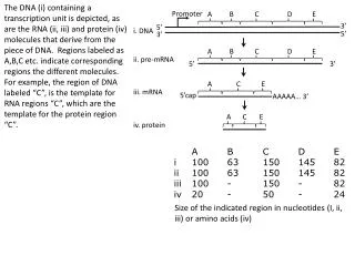
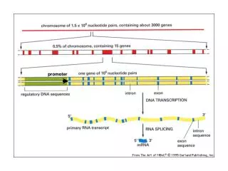
Prokaryotic Genes Translational start site (AUG) Translational stop site Promoter region Open Reading Frame Transcriptional start site Operator sequence Transcriptional stop site Recall - prokaryotes have a single circular chromosome Also, no cell nucleus, and no introns Therefore, prokaryotic gene structure is quite simple
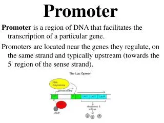
Prokaryotic Operons Operon structure Upstream Downstream Promoter Gene 1 Gene 2 Gene 3 In prokaryotes, sometimes genes that are part of the same operational pathway are grouped together under a single promoter. They then produce a pre-mRNA.
Bacterial Gene: Signals +1 Gene 2 Gene 1 Bacterial genomes have simple gene structure • - Promoter • -35 sequence (T82T84G78A65C54A45) 15-20 bp • -10 sequence (T80A95T45A60A50T96) 5-9 bp (Pribnow Box) • Start of transcription : +1 initiation start: Purine90 • Translation binding site (Shine-Dalgarno) 10 bp upstream of • AUG (AGGAGG) • - One or more Open Reading Frame • Start-codon: ATG (unless sequence is partial) • stop codon for gene 1 .. • Separated by intercistronic sequences.
Promoters • Promoter sequences facilitate the binding of the RNA polymerase to the DNA to be transcribed. • Promoters of different genes have distinct sequences, although most have characteristic short sequences of 6 to 10 bases at a position between 10 to 30 nucleotides upstream -10 sequence: Hexamer: TATAAT– Pribnow Box (Pribnow, 1975) and -35 sequence, an hexamer : TTGACA in prokaryotes.
Typical E. coli Promoters TATAAT Pribnow Box
mRNA start point -35 region Pribnow box ‘Consensus’ sequences of E. coliPromoters T80A95T45A60A50T96 • the sequence at the promoter can regulate efficiency of initiation • different sigma factors may associate with RNA polymerase, which target specific promoters
Methods for characterization of promoters DNAse protection method DMS protection method Foot-printing method
1. DNase Protection method The region of DNA in contact with RNA polymerase can be isolated • Allow the piece of DNA containing the promoter to interact with RNA polymerase • Treat with DNase I • Dissociate the enzyme and isolate the DNA • Determine the size by gel electrophoresis • Determine the sequence by standard method
_ _ _ _ _ _ _ _ _ _ _ _ DNase I + Mono and dinucleotides DNA molecule with promoter RNAP + DNA complex RNAP Dissociate DNA from enzyme Sequence the Promoter DNA Promoter region
2. DMS Protection method Specific points of contact within the contact region can be identified Dimethyl sulphate methylates N3 of A or N7 of G, but not C or T Glycosidic bond of methylated As or Gs is unstable and can be broken by heating at neutral pH leaving deoxy ribose from the chain – DNA degradation results Region of the DNA bound by RNA polymerase will not be methylated – it will be intact Dissociate the enzyme and isolate the DNA fragment that corresponds to the promoter
DNA molecule with promoter RNAP DMS _ _ _ _ _ _ _ _ _ _ _ _ * * * * * + * * * * * Mono and dinucleotides RNAP + DNA complex Methylated purines Dissociate DNA from enzyme Sequence the Promoter DNA Promoter region
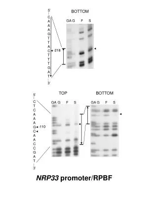
3. Foot-printing method • Take a DNA fragment with known Restriction sites • Dephosphorylation – Alkaline phosphatase • End labelling – 5’ is to be labelled with • gamma-32P- ATP using T4 polynucleotide kinase • Remove a small fragment by RE digestion • Allow the labelled DNA to interact with RNA polymerase • - One sample is to be maintained without RNAP • treatment • Using DNA endonuclease briefly digest the DNA sample treated with RNAP – Nicking occurs randomly at all places except those protected by RNAP • Analyze both the samples (with and without RNAP interaction) following agarose gel electrophoresis A method to detect where a protein binds to DNA
Foot-printing One end labelled DNA RNAP Used extensively for mapping contact points between promoter sequences and RNA polymerase and/or regulatory proteins No RNAP
Interpretation If the DNA contains ‘n’ bp and RNAP is not added, ‘n’ sizes of DNA fragments will be present However, if RNAP binds to ‘x’ bp and thereby prevents access of the DNA to the nuclease, only ‘n–x’ different sizes of DNA fragments will be represented The positions of the missing bands are the positions of the ‘n’ bands on DNA