Download

1 / 74
830 likes | 1.32k Views
Genitourinary trauma. Prepared by : Hashim Gulam , R2. introduction. Traumatic injury is a leading national and international health problem. In the United States, 1 of every 14 deaths—over 150,000 per year—results from trauma.

E N D
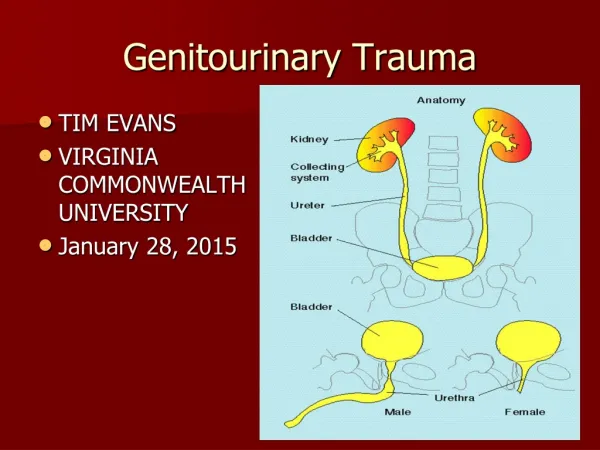
Genitourinary trauma Prepared by:Hashim Gulam , R2
introduction • Traumatic injury is a leading national and international health problem. • In the United States, 1 of every 14 deaths—over 150,000 per year—results from trauma. • trauma results in more deaths between ages 1 and 37 years than any other cause.
Gu trauma • Renal Trauma • Ureteric Trauma • Bladder Trauma • Urethral Trauma
RENAL TRAUMA • Of all injuries to the genitourinary system, injuries to the kidney from external trauma are the most common (50% of all genitourinary trauma). Mechanism of injury: • Blunt (motor vehicle accident, assault, falls) • Penetrating ( gunshot wounds, stab wounds) • Iatrogenic ( endourologicprocedures,ESWL, renal biopsy, percutaneous renal procedures)
Clinical Diagnosis • Mechanism of injury provides the framework for clinical assessment. • Hematuria is the best indicator of traumatic urinary system injury. • The degree of hematuria doesn’t correlate with the degree of injury. • Flnk pain & tenderness,fractures of the lower ribs and upper lumbar and lower thoracic vertebrae are associated with renal injuries.
Indications for Renal Imaging • Blunt trauma patients with gross hematuria • Patients with microscopic hematuria and shock (systolic blood pressure of less than 90 mm Hg any time during evaluation and resuscitation) • Penetrating injuries with any degree of hematuria • Pediatric trauma patient with gross or significant microscopic hematuria (>50 RBC/HPF) • Associated injuries suggesting underlying renal injury • Major deceleration injury
Imaging Studies • The preferred imaging study for renal trauma is contrast-enhanced CT. • It is Highly sensitive and specific. provides the most definitive staging information: • parenchymal lacerations, extravasation of can easily be detected & associated injuries to the abdominal organs can be identified.
the degree of retroperitoneal bleeding can be assessed by the size and dimensions of the retroperitoneal hematoma. • Lack of uptake of contrast material in the parenchyma suggests arterial injury.
IVP: • All patients who require immediate surgical exploration should undergo a one-shot high dose IVP (2 mls\kg of 60% contrast followed by a single KUB 10 minutes later). Angiography: • to define arterial injuries suspected on CT or to localize arterial bleeding that can be controlled by embolization
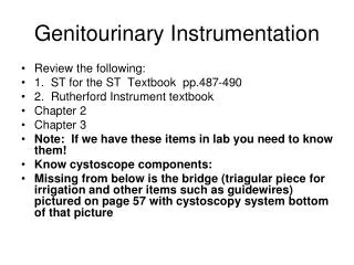
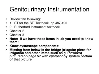
right renal stab wound (grade IV), demonstrating extensive urinary extravasation and large retroperitoneal hematoma
Delayed imaging Injury to collecting system with extravasation
Delayed imaging Renal pelvis injury with leak of urine
management Non operative: • Majority of renal injuries can be managed conservatively. • Admission to the hospital & bed rest till the urine become clear. • Close monitoring of vitals & serial Hb, Hct. • Close clinical follow up after discharge.
Indication for renal exploration Absolute: • Persistent renal bleeding • Pulsatile,expanding or uncontained hematoma • Avulsion of the main renal artery or vein
Relative: • Significant (25%-50%) non-viable tissue. • Urinary extravasation . • Arterial thrombosis. • Penetrating trauma.
Renal exploration • The goals of operative therapy are hemorrhage control and renal tissue preservation • Surgical exploration of the acutely injured kidney is best done by a transabdominal approach, which allows complete inspection of intra-abdominal organs and bowel.
Renal exploration, débridement of nonviable tissue, hemostasis by individual suture ligation of bleeding vessels, watertight closure of the collecting system, and coverage or approximation of the parenchymal defect
When polar injuries cannot be reconstructed, a partial nephrectomy should be done and all nonviable tissue removed, hemostasis obtained, and the collecting system closed. The open parenchyma should then be covered when possible by a pedicle flap of omentum.
Indication for nephrectomy • Total nephrectomy would be indicated immediately in extensive renal injuries when the patient's life would be threatened by attempted renal repair. • Grade 5 injuries that deemed irreparable. • Major vascular injury.
Complications • Urinoma: treated by systemic antibiotics if persist, internal ureteric stent often correct the problem. • Delayed renal bleeding: occur several weeks after injury &The initial management is bed rest and hydration & if persist, angiography and embolization can often gain control. • Perinephric abscess: treated by percutaneous drainage. • HTN
Ureteral injury • Ureteral injuries after external violence are rare, occurring in less than 4% of cases of penetrating trauma and less than 1% of cases of blunt trauma. • Majority of ureteral injury are iatrogenic injuries. • hysterectomy responsible for the majority of ureteral injury(54%), followed by colorectal surgery (14%), pelvic surgery such as ovarian tumor removal (8%), and abdominal vascular surgery (6%)
diagnosis • Intra-operative recognition. • 70-80% of iatrogenic injuries are diagnosed postoperatively. • The presenting signs and symptoms may include flank pain (36-90%), fever and sepsis (10%), fistula, urinoma, prolonged ileus, and renal failure from bilateral obstruction (10%).
Imaging study • IVP • CT • Retrograde pyelogram • Antegrade pyelogram
management External trauma Contusion: • if minor, stent placement. • if large contusion, excision &U-U upper ureteral injury: • direct u-u • transureteroureterostomy • autotransplantation • ileal interposition segment: only for delayed repair
midureteral injury: • direct u-u • trans u-u lower ureteral injury: • uretericreimplantation • psoas hitch. • Boari flap.
Suggested management options for ureteral injuries at different levels.
Technique of ureteroureterostomy after traumatic disruption: A, injury site definition by ureteral mobilization; B, débridement of margins and spatulation; C, stent placement; D, approximation with 5-0 absorbable suture; E, final result
Transureteroureterostomy • The donor ureter is brought to the contralateral side through a tunnel under the mesentery of the sigmoid colon superior to the inferior mesenteric artery. • The anastomosis is performed in an end-to-side fashion.
Absolute contraindications to this procedure include a short donor ureter or a diseased recipient ureter • Relative contraindications include a urothelial tumor, nephrolithiasis, pelvic or abdominal irradiation, retroperitoneal fibrosis, and in cases of a ureteral injury during aortoiliac bypass surgery
ureteroneocystostomy • Ureteroneocystostomy is used to repair distal ureteral injuries that occur so close to the bladder that the bladder does not need to be brought up to the ureteral stump with a psoas hitch or Boari procedure.
psoas hitch • Used if a tension-free anastomosis cannot be accomplished by a simple ureteroneocystostomy. • Can be used to bridge a 6- to 10 cm defect . • Involves mobilizing the bladder and pulling it superiorly and laterally by fixing it to the psoas tendon with absorbable suture. • Care must be taken to avoid injuring the genitofemoral nerve
Boari bladder flap • It involves creating a posterolateral bladder flap based on the superior vesical artery or one of its branches. • Used in Injuries to the lower two thirds of the ureter with long ureteral defects. • The procedure is time consuming & not commonly used
Surgical injury: • Ligation: removal of the ligature and observation of the ureter for viability. If viability is in question, ureteroureterostomy or ureteralreimplantation should be performed. • Stenting is advised.
Transection: • Early recognition:nephrectomyvs U-U. • Delayed recognition:stent placement for 6w-3m. • If failed, place NT & attempt stenting after 2w • wait several months and perform open repair in pts w/ persistent leaks or ureteral stricture
Ureteroscopy Injury: • Avulsion: treated in the same manner as ureteral injuries after open or laparoscopic surgery. • Perforation: treated by ureteralstenting, usually with no subsequent complications.
Bladder injury Etiology: • Blunt: MVA, assault, falls. • Penetrating:gunshot wound, stab wound. • Iatrogenic: obstetric, gynecological, urological & orthopedic procedure. • The most common associated injury is pelvic fracture, associated with 83% to 95% of bladder injuries.
classification Bladder contusion: trauma pt w/ hematuria w/ no evidence of urethral or renal injury and normal cystogram. Extraperitoneal bladder rupture: with pelvic # Intraperitoneal bladder rupture: with penetrating inj. Combination of intraperitoneal and extraperitoneal ruptures
Clinical presentation • A triad of symptoms is often present (gross hematuria, suprapubic pain or tenderness, difficulty or inability to void) • An abdominal examination may reveal distention, guarding, or rebound tenderness • Absent bowel sounds and signs of peritoneal irritation indicate a possible intraperitoneal bladder rupture
Bilateral palpation of the bony pelvis may reveal abnormal motion indicating an open-book fracture or a disruption of the pelvic girdle. • If blood is present at the urethral meatus, suspect a urethral injury.