Download
1 / 24
240 likes | 387 Views
HEMATURIA BASIC COURSE OF DIAGNOSIS. Xiaoqi Xu Renji Hospital Shanghai Second Medical University. CONTENT. Definition of hematuria Etiology Clinical feature Differential diagnosis Laboratory tests Accompanied symptoms. DEFINITION.

E N D
HEMATURIABASIC COURSE OF DIAGNOSIS Xiaoqi Xu Renji Hospital Shanghai Second Medical University
CONTENT • Definition of hematuria • Etiology • Clinical feature • Differential diagnosis • Laboratory tests • Accompanied symptoms
DEFINITION More than three red blood cells are found in centrifuged urine per high-power field microscopy ( > 3 RBC/HP). Normal urine: no red blood cell or less than three red blood cell
According to the amount of RBC in the urine, hematuria can be classified as: • microscopic hematuria: normal colour with eyes • gross hematuria: tea-colored, cola-colored, pink or even red
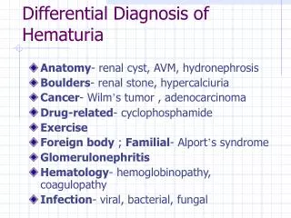
ETIOLOGY • Diseases of the urinary system—the most common cause Vascular arteriovenous malformation arterial emboli or thrombosis arteriovenous fistular nutcracker syndrome renal vein thrombosis loin-pain hematuria syndrom cogulation abnormality excessive anticogulation
Glomerular IgA nehropathy thin basement membrane disease (incl.Alport syndrome) other causes of primary and secondary glomerulonephritis Interstitial allergic interstitial nephritis analgesic nephropathy renal cystic diseases acute pyelonephritis tuberculosis renal allograft rejection
Uroepithelium malignancy vigorous excise trauma papillary necrosis cystitis/urethritis/prostatitis(usually caused by infection) parasitic diseases (e.g. schistosomiasis) nephrolithiasis or bladder calculi Multiple sites or source unknown hypercalciuria hyperuricosuria
System disorders a. Hematological disorders aplastic anemia leukemia allergic purpura hemophilia ITP (idiopathy thrombocytopenic purpura) b. Infection infective endocarditis septicemia epidemic hemorrhagic fever (Hantaan virus) scarlet fever (-hemolytic streptococcus) leptospirosis (leptospire) filariasis (Wuchereria bancrofti, Brugia malayi)
c. Connective tissue diseases systemic lupus erythematosus (SLE) polyarteritis nodosa d. Cariovascular diseases hypertensive nephropathy chronic heart failure renal artery sclerosis e. Endocrine and metabolism diseases gout diabetes mellitus
Diseases of adjacent organs to urinary tract appendicitis salpingitis carcinoma of the rectum carcinoma of the colon uterocervical cancer • Drug and chemical agents sulfanilamides anticogulation cyclophosphamide mannitol • miscellaneous exercise “idopathic” hematuria
CLINICAL FEATURE • Color depends on the amount of red blood cell in the urine and the pH (see slide 4) normal: light yellow, pH 6.5 • pH acidic: more darker (brown or black) alkaline: red
DIFFERENTIAL DIAGNOSIS • Polluted urine: menstruation • Drug and food: phenosulfonphtha lein (PSP),uric acid, vegetable • Porphyrism: porphyrin in urine (+) • Hemoglobinuria hemolysis soy-like, very few RBC under the microscopy occult blood test (+)
HEMOGLOBINURIA RBC abnormality • Defects of RBC membrane structure and function (hereditary spherocytosis) • Deficiency of enzymes (favism) • Hemoglobinopathy (thalassemia) • PNH Mechanical factor (artificial heart valve), infection or mismatched blood transfusion
LABORATORY TESTS • Three-glass test Method: collecting the three stages of urine of a patient during micturition Result: • the initial specimen containing RBC—the urethra • the last specimen containing RBC—the bladder neck and trianglar area, posturethra • all the specimens containing RBC—upper urinary tract, bladder
Phase-contrast microscopy to distinguish glomerular from post glomerular bleeding • post glomerular bleeding: normal size and shape of RBC • glomerular bleeding: dysmorphic RBC (acanthocyte)
EXAMPLE OF PHASE-CONTRAST MICROSCOPY TEST (non-glomerlar) RBC MCV: 92.8 um3
ACCOMPANIED SYMPTOMS • Hematuria with renal colic renal stone, ureter stone if with dysuria, miction pause or staining to void: bladder or urethra stone • Hematuria with urinary frequency,urgency and dysuria bladder or lower urinary tract (tuberculosis or tumor) if accompanied by high spiking fever, chill and loin pain: pyelonephritis
Hematuria with edema and hypertension glomerulonephritis hypertensive nephropathy • Hematuria with mass in the kidney neoplasm hereditary polycystic kidney • Hematuria with hemorrhage in skin and mucosa hematological disorders infectious diseases • Hematuria with chyluria filariasis
--Approaching to the patient– (Harrison’s Principle of Internal Medicine,14th Ed) HEMATURIA proteinuria (>500mg/24h) Dysmorphic RBC or RBC casts (-) (+) (+) Pyuria,WBC casts urine culture eosinophils serologic and hematologic evaluation: blood culture, anti-GBM Ab, ANCA, complement, cryoglobulin HBV,HCV,VDRL,HIV, ASLO (-) Hb electrophoresis, urine cytology, UA of family member, 24h urinary calcium/uric acid (-) As indicated: retrograde pyelography or arteriogram of cyst aspiration (+) IVP+/-renal ultrasound renal biopsy (-) (+) cystoscopy biopsy (-) ANCA:antineutrophil cytoplasmic antibody, VDRL:venereal dis. research laboratory, ASLO: antisteptolysin O, IVP: intravenous pyelography CT scan (+) open renal biopsy (-) follow