Download
1 / 22
220 likes | 361 Views
Morning Report 7/31/07 . 3 rd Degree AV block Jason Haag. Heart Block. 1 st Degree AV Block one-to-one relationship exists between P waves and QRS complexes, but the PR interval is longer than 200 ms. Heart Block. 2 nd Degree Mobitz Type I AV Block (Wenckebach)

E N D
Morning Report7/31/07 3rd Degree AV block Jason Haag
Heart Block • 1st Degree AV Block • one-to-one relationship exists between P waves and QRS complexes, but the PR interval is longer than 200 ms
Heart Block • 2nd Degree Mobitz Type I AV Block (Wenckebach) • PR interval is prolonging with each P wave to the point when the P wave is no longer conducted
Heart Block • 2nd Degree Mobitz Type II AV Block • PR interval is constant, but occasionally P waves are not followed by the QRS complexes
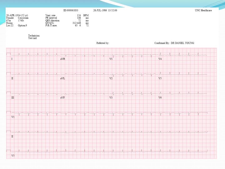
Heart Block • 3rd Degree Heart Block • More P waves than the QRS complexes exist and no relationship exists between them
3rd Degree Heart Block • Block can be in AV node or infranodal conduction system • AV node • 2/3 escape rhythms have narrow QRS (junctional) • Fascicular or bundle branches • Wide QRS (subjunctional) • Rate typically in low 40s
Frequency • In the US: 0.02% • Internationally: 0.04%. • Age: Bimodal peak, at infancy given congenital complete AV block and at advance d age due to progressive fibrosis and ischemia
History • Syncope, near-syncope, and lightheadedness • Fatigue, dyspnea, and angina • Asymptomatic • Sudden cardiac death
Physical • Vital Signs (stable vs. unstable, always check HR manually) • Signs of heart failure – JVD, a waves, Pulmonary edema • New murmurs or gallops • Target lesions (Lyme) • Splinter hemm, Osler nodes, etc (endocarditis) • Neuromuscular changes (mytonic/muscular dystrophy)
Etiologies • Idiopathic Progressive Cardiac Conduction Disease • ½ of cases of AV block • Lenegre’s disease • Progressive, fibrotic, sclerodegeneration of the conduction system • Younger individuals, may be hereditary • Lev’s disease • Calcification extending from fibrous structures (aortic/mitral rings) into the conduction system • Older individuals, ? ESRD • Fibrosis NOS • Typically mitral and aortic rings • Mitral narrow QRS • Aortic wide QRS
Etiologies (cont.) • Ischemic heart disease • 40% of cases • Either from chronic ischemia or acute MI • Acute MI AV blocks (20% of patients) • 1st degree (8%) • 2nd degree (5%) • 3rd degree (6%) • LBBB/RBBB (10-20%) • AV nodal block (narrow QRS) associated with inferior wall MI • Bundle blocks (wide QRS) associated with anterior wall MI • Drugs • Calcium channel blockers, beta blockers, digoxin, amiodarone, adenosine, quinidine, procainamide
Etiologies (cont.) • Infection • Lyme disease, endocarditis, Rheumatic fever, Chagas disease, myocarditis • Rheumatic disease • Ankylosing spondylitis, Reiter syndrome, relapsing polychondritis, rheumatoid arthritis, scleroderma • Infiltrative disease • Amyloidosis, sarcoidosis, multiple myeloma, hemachromatosis, Wilson’s disease
Etiologies • Hyperthyroidism • Metabolic • Hypoxia, hyperkalemia • Neuromuscular disease • Muscular dystrophy, dermatomyositis
Treatment • Correct underlying problem – if you can • Correct K, stop AV blocking medications, etc. • If unstable • Transcutaneous pacing • If stable • Plan for permanent pacemaker placement
Permanent Pacemaker • Class I - Conditions for which evidence and/or general agreement exists that a given procedure or treatment is beneficial, useful, and effective • Third-degree AV block and advanced second-degree AV block at any anatomic level associated with any one of the following conditions: • Bradycardia with symptoms, heart failure, arrhythmias, pauses greater than 3 seconds, escape rate < 40 bpm
Permanent Pacemaker • Class IIa - Weight of evidence or opinion is in favor of usefulness or efficacy • Asymptomatic third-degree AV block at any anatomic site with average awake ventricular rates of 40 bpm or faster, especially if cardiomegaly or left ventricular (LV) dysfunction is present
References • Gregoratos G, Abrams J, Epstein AE, et al: ACC/AHA/NASPE 2002 guideline update for implantation of cardiac pacemakers and antiarrhythmia devices: summary article: a report of the American College of Cardiology/American Heart Association Task Force on Practice Guidelines. Circulation 2002 Oct 15; 106(16): 2145-61. • Kojic EM, Hardarson T, Sigfusson N, Sigvaldason H: The prevalence and prognosis of third-degree atrioventricular conduction block: the Reykjavik study. J Intern Med 1999 Jul; 246(1): 81-6. • McEnvoy GK, ed: AHFS Drug Information 2000. Bethesda, Md: American Society of Health-System Pharmacists; 2000: 1187-95. • Ostaner LD, Brandt RL, Kjelsberg MI, et al: Electrocardiographic findings among the adult population of a total natural community. 1965; 31: 888-98. • Rardon DA, Miles WM, Mitrani RD, et al: Electrocardiographic Recognition: Atrioventricular Block and Dissociation. In: Zipes DP, Jalife J, eds. Cardiac Electrophysiology From Cell to Bedside, 2nd ed. Philadelphia, Pa: WB Saunders; 1995.