Download

1 / 82
870 likes | 1.15k Views
Laboratory Methods for Diagnosis of Non-fermenting Gram-Negative Bacilli. Dr Mohammad Rahbar. General Characteristics of Non-fermenters.

E N D
Laboratory Methods for Diagnosis of Non-fermenting Gram-Negative Bacilli Dr Mohammad Rahbar
General Characteristics of Non-fermenters • Nonfermenting gram-negative bacilli are grouped together because they fail to acidify oxidative-fermentative (OF) media overlaid with mineral oil or triple sugar iron agar (TSIA) butts .They prefer and grow much better in an aerobic environment ;some group members oxidize carbohydrates to derive energy for their metabolism ;they are referred to as oxidizers.
General Characteristics of Non-fomenters • Others do not break down carbohydrates at all and are inert or biochemically inactive; they are referred to as nonoxidizer or asaccharolytic .Additional characteristics can differentiate this group of nonfernenters from other gram-negative bacilli: motility ,pigmentation and their ability or lack of ability to grow on selective gram-negative media such as MacConkey agar.
General Characteristics of Non-fermenters • Most nonfermentative gram-negative bacilli are oxidase positive, a feature that differentiate them from the Enterobacteriaceae (except plesiomonas witch is oxidase positive.
General Characteristics of Non-fermenters • In general nonfermentative gram-negative bcilli are ubiquitous and found in most environments: in soil and water .on plants and decaying vegetation and in many foodstuffs. They prefer moist environment ,and in hospitals that can be isolated from nebulizers, dialysate,fluide saline and on catheters and other devices.
General Characteristics of Non-fermenters • Nonfermenters may withstand treatment with chlohexidine and quaternary ammonium compounds .They are rarely ,if ever part of the normal host flora but can easily colonize hospitalized patients, especially those who are immunocompromised .Nonfermentative gram-negative bacilli tend to be resistant to several Antimicrobial agents
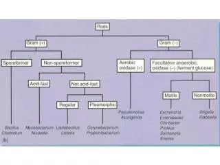
TAXONOMY, BIOCHEMICAL CHARACTERISTICS, AND CLINICAL SIGNIFICANCE OF MEDICALL Y IMPORTANT GENERA OF NONFERMENTERS
Continue.. • Unlike the Enterobacteriaceae the nonfermenting gram-negative bacillido not fit conveniently into a single family of well-characterized genera, and the correct taxonomic placement of many nonfermentative, gram-negative bacilli (NFBs) remains unresolved.
Continue… Consequently, the study of nonfermenters is often confusing for the beginning microbiologist. The major genera of nonfermenting, gram-negative bacilli have been classified into at least 15 families.
Continue… • One approach to studying the nonfermenters is to group them on the basis of the presence or absence of motility and on the type of flagella present in strains that are motile.
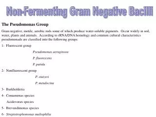
Continue… • Organisms That Are Motile With Polar Flagella Pseudomonads
Fluorescent Group. • The species within this group are all by the production of a water-soluble white to blue-green under long wavelength pyoverdin pigment that fluoresces (400-nm) ultraviolet light. Production of fluorescent pigments is particularly enhanced in media with a high phosphate concentration.
Continue… • Although all three members of this group produce pyoverdin, only one species. P. aeruginosa ,produces the distinctive blue, water-soluble pigment pyocyanin
Continue… • Pseudomonas aeruginosa produces a characteristic appearance when grown on BAP. It appears as large gray colonies with a spreading periphery and exhibits hemolysis. Colonies often have an alligator skin appearance and exhibit a metallic sheen.
Continue… • Rapid identification of P. aeruginosa in culture can be made whenever the characteristics are observed: typical colony morphology production of diffusible pigmentsthe presence of a fruity odor, and oxidase positivity .
Continue…. We have occasionally observed strains that produce a pungent, "rotten-potato" odor. There has been at least one report of a nosocomial outbreak caused by strains of malodorous P. aeruginosa .
Continue… • Pseudomonas aeruginsa is the most frequently recovered from clinical specimens. p, aeruoginosa infection is especially prevalent among patients with burn wounds, cystic fibrosis, acute leukemia, organ transplants, and drug addiction.
Continue… • Infections commonly occur at any site where moisture tends to accumulate tracheostomies, indwelling catheters, burns, the external ear ("swimmer's ear"), and weeping cutaneous wounds. The exudation of bluish pus, with a grape-like odor from the production of pyocyanin, is characteristic.
Continue • P aerugnosa also causes urinary tract and lower respiratory tract infections; the latter can be severe and even life-threatening in immunocompromised hosts. The organism can also cause devastating infections of the eye
Continue… • Pseudomonas keratitis. Infection of corneal ulcers, and endophthalmitis must be approached as a medical emergency that can be fulminant and threaten permanent loss of vision. Individual cases of endocarditis,meningitis, brain abscess, and infections of bones and joints from hematogenous spread appear with regular frequency in the literature.
Continue.. • Most cases of endocarditis require valve replacement because the infection is difficult to eradicate. P. aeruginosadermatitis and otitis externa outbreaks associated with swimming-pool and hot-tub use are well described. The CDC reported at least 75 cases during six outbreaks occurring between 1997 and 1998. Sporadic P. aeruginosa infections following ear piercing have also been reported.
Continue… • P .aerugillosa produces several substances that are thought to enhance the colonization and infection of host tissue. These substances, together with a variety of virulence factors, including lipopolysaccharide (LPS), exotoxinA, leukocidin, extracellular slime, proteases, phospholipase,and several other enzymes (Box 7-5), make P. aerugillo.l'a the most clinically significant bacteria among the NFB.
Virulence Factors • An unusual mucoid morphotype of P. aeruginosa is frequently recovered from respiratory secretions of patients with cystic fibrosis who are chronically infected with P. aeruginosa The mucoid morphotype is due to the production of large amounts of a (called alginate)that surrounds the cell. The production of alginate is ultimately responsible for the poor prognosis and high mortality rates among patients with cystic fibrosis.
Virulence Factors of Pseudomonasaeruginosa • Alginate: Capsular polysaccharide that allows infecting bacteria to adhere to lung epithelial cell surfaces and form biofilms which, in turn, protect the bacteria from antibiotics and the body's immune system
Virulence factors • PIlli Surface appendages that allow adherence of organism to GM-I ganglioside receptors on host epithelial cell surfaces Neuraminidase Removes sialic acid residues from GM-I ganglioside receptors. Facilitating binding pili
Viurlence factors • Exotoxin A : • Tissue destruction, inhibition of protein synthesis; interrupts cell activity . Enterotoxin • Interrupts normal gastrointestinal activity. leading to diarrhea
Virulence Factors • Exoenzyme S: Inhibits protein synthesis Phospholipase C: Destroys cytoplasmic pulmonary surfactant; inactivates opsonins
Virulence Factors • Elastase: Cleaves immunoglobulins and , disrupts neutrophil activity • Leukocidin: • Inhibits neutrophil and lymphocyte function
Virulence Factors • Pyocyanins: • Suppress other bacteria and disrupt ciliary activity; cause oxidative damage to tissues, particularly oxygenated tissues such as lung
Summary Key Tests for Identification P. aeruginosa • Minimum Requirements for Definitive Identification of P. aeruginosa Identification based on all of the following: • I. Gram-negative rod • 2. Oxidase-positive • 3. Typical smell (fruity grape-like odor or corn tortilla) • 4. Recognizable colony morphology • a. On blood or chocolate agar appear as large colonies with • metallic sheen, mucoid, rough. or pigmented (pyocyanin) • and often p-hemolytic
Summary Key Tests for IdentificationP. aeruginosa b. On MacConkey, appear as lactose-negative with greenpigmentation, or metallic sheen Limitations: I. Rare Aeromonas isolates may resemble P. aerugirrosa (lacking the typical smell) but will be spot indole-positive (P. aeruginosq are indole-negative). 2. Some Burklto/der;a cepac;a isolates from patients with cystic fibrosis may exhibit morphotypes that resemble P.aeruginosa.
Colonies of Pseudomonas aeruginosa typically display beta hemolysis, a metallic sheen, and blue or green pigment.
Pseudomonas aeruginosa (beta hemolysis with transmitted light)
. Pseudomonas aeruginosa (beta hemolysis with transmitted light
FIG. 5. Pseudomonas aeruginosa (beta hemolysis and pigment with transmitted light
encapsulated strain of Pseudomonas aeruginosa recovered from a cystic fibrosis patient at 24 hours.
Same plate as FIG. 23 at 48 hours, this strain of Pseudomonas aeruginosa make abundant, mucoid capsular material.
Acinetobacter • The genus Acinetobacter ,now a member of the family Moraxellaceae ,cosist of 25 ِDNA homology groups or genomospeecies .Only 10 species have been officially named:the two species most commonly seen in clinical specimens are : A.baumannii and A. lwoffii
Continue.. • Acinetobacter spp are unique in the environment in soil, water and foodstuffs in the hospital environment they have been associated with ventilator ,humidifies catheter and other devices. About 25% of adults carry the organism in their phrynx.If not harboring Acinetobacter spp ,already hospitalized patients may become easily colonized,
Continue.. • As many as 45% of patents with a trachetomy may be colonized. When Acinetobacter spp isolated from urine, feces ,vaginal secretion ,and many different type of respiratory specimens, they are often considered insignificant colonizer or contaminants.
Acinetobacter baumannii • A. baumannii is the second most frequentnonfermenter encountered in clinical laboratories, but with only about one tenth the frequency of P. aerugi1losa. The following are the characteristics by which a presumptive identification can be made.
Clinical Infections • Acinetobacter spp are opportunistic accounting 1% to 3% of all nosocomial infections< they are second only to P.aeruginosa in frequency of isolation of all nonfermenters in the clinical microbiology laboratory.
Disease in particular with A.baumannii • UTI • Pneumonia, Tracheobronchitis,or both • Endocarditis with up 25% mortality • Meningitides • SepticemiaTruman infections, Burn infections, • Eye infections. • A.lwoffii is much less virulent
Laboratory Diagnosis • Appear as cocci or coccobacilli on Gram stain . • Grow well on MacConkey agar (colonies may have slightly pinkish tint ,a helpful characteristic when present • Exhibit rapid utilization of glucose, with production of acid • Are non- motile • Are penicilin resistant
Lab Diagnosis • The initial clue is the observation of tiny diplococci on Gram stains prepared directly from clinical materials. When Gram stains are prepared from agar or broth cultures, the cells may appear larger and more like coccobacilli
Lab Diagnosis • Acinetobacter species are not pigmentedwhen grown on blood agar, a helpful characteristic in differentiating them from certain other nonfermenters, such as occasional oxidase-negative, nonmotile strains of Burkholderia cepacia.
Lab Diagnosis • However, colonies growing on Mac- Conkey agar may produce a faint pink tint or a deeper cornflower blue when observed on eosin methylene blue agarResistance to penicillin helps distinguish • A. baumannii from the highly penicillin-sensitive Moraxel/ a species, which also usually appear as coccobacilli on Gram stain.
Lab Diagnosis • Most strains of Moraxel/a species are also cytochrome oxidase-positive. A. lwoffii is nonsaccharolytic and can be differentiated from A. baumannii because it produces no acid when grown in media that contain carbohydrates.


![GRAM NEGATIVE BACILLI- MICRO {ST1]](https://cdn1.slideserve.com/2240310/gram-negative-bacilli-micro-st1-dt.jpg)