Download

1 / 26
260 likes | 398 Views
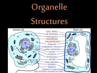
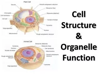
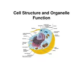
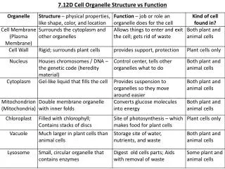
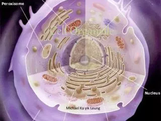
Organelle structure. Nucleus Sarco(endo)plasmic reticulum Mitochondria Peroxisome Autophagosome. Electron microscope. Diffraction limit Wave nature of light passing through lens Optical: l ~0.6 um, resolution ~0.2 um Confocal, FT, nanohole arrays De Broglie wavelength l = h/mv

E N D
Organelle structure • Nucleus • Sarco(endo)plasmic reticulum • Mitochondria • Peroxisome • Autophagosome
Electron microscope • Diffraction limit • Wave nature of light passing through lens • Optical: l~0.6 um, resolution ~0.2 um • Confocal, FT, nanohole arrays • De Broglie wavelength • l= h/mv • Electron: l = 1.2(V- ½) nm (V: accelerating voltage) B~l/(2 NA)
EM imaging • Essentially a CRT • Vacuum • Focus e-beam on specimen • Expand onto screen • Sample prep • Dehydrate & fix • Heavy metal stain (uranium) • 90 nm slices (Graham Colm, wikipedia)
Structure Nucleus Myofibril Artifact Mitochondria Out of plane
Common features • Densely packed myofibrils • Subsarcolemmal space • Nuclei • Mitochondria • Intermyofibrillar I-band space • Mitochondria • SR/T-tubule triads
Fractional composition • Stereology • Statistical model of 3-D structure • 2-D images & discrete sampling
Rattlesnake tail muscle: prominent extensive glycogen & mitochondria; relatively few myofibrils Clark & Schultz, 1980
Nucleus • Multiple • MND 3e4-5e4 um3/nucleus • Fiber volume 5e5-5e6 um3 (10-1000 nuclei) • Cylindrical, ~30 um3 • Nucleolus • Cajal bodies • DNA • RNA processing Terada et al., 2010
RNA processing • Nucleolus: rRNA synthesis & ribosome assy • Cajal bodies: Spliceosome assembly • Speckles: RNA splicing Pollard & Earnshaw, 2008
Ribosomal biogenesis • Multiple (300+) copies of rRNA strand • Transcribed • Cleaved to 28S, 18S, 5.8S strands • Ribose methylation • Uridine isomerization to pseudouridine • Ribosomal proteins • Imported to nucleus • Anneal to rRNA in nucleolus
Nuclear Envelope • Dual membrane • Control nuclear import/export • Nuclear pore complex
Nuclear membrane proteins • Chromatin anchors • Lamin, lamin B receptor, emerin • Transport proteins • Importin, Exportin, Ran
Mitochondria • Dual membrane • Oxidative ATP synthesis • Citric acid cycle • Electron transportchain • Separate genome • Most Mt proteins come from nuclearDNA
Mitochondrial syncytium • Network, not bacterium-like • Regulated growth • Division • Fusion • Apoptosis Manella, 2000
Muscle mitochondria • Extensive subsarcolemmal syncitia • Elongated tubules, perpendicular to fiber • Frequently doubled around Z-disks Subsarcolemmal mitochondria Intermyofibrillar mitochondria Ogata & Yamasaki, 1997
Inner membrane • Cristae • “Shelf-like” • Tubular network Manella, 2008
Electron transport chain NADH FADH2 Ubiquinone Cytochrome C Reactive oxygen Oxidative ATP Synthesis
Peroxisome • Single membrane • Controlled H2O2 chemistry • Oxidases generate H2O2 • Peroxidases degrade H2O2 • Fatty acid metabolism • Cholesterol & bile synthesis • Peroxins (PEX) control protein composition and biogenesis
Sarcoplasmic Reticulum • Single membrane • Lamellar, fenestrated • Terminal cisternae • Synthesis of transmembrane and extracellular proteins • Calcium storage
SR T-tubule communication • Terminal cisternae wrapT-tubule • Communication of membrane events to SR SEM of I-band region of a myofiber with myofibrils extracted, leaving only membrane-bounded skeletons of T-tubule (T), SR (blue), terminal cisternae (arrowheads), and mitochondria. Fiber axis is vertical. Ogata & Yamasaki, 1997
Triad • Physical association between T-Tubule and SR • “Feet” • Dihydropyridine receptor (DHPR) • Ryanodine Receptor (RyR) Franzini-Armstrong, 1970
“white” vs “red” muscle • Red: larger, more extensive mitochondria • White: (slightly) more extensive SR
Autophagosome • Double membrane • Small (1 um), spherical • Self-engulfment • Recruit lysosome & degrade Mizushima & al., 2002
Autolysis • Degradation of large structures • Starvation defense • Infection defense • Targeting • Apg5/Apg12 targeting complex • LC3 microtubule associated light chain • PI3k, FOXO3
Summary • Specialized chemistry is constrained to isolated cellular domains • Nucleus • Mitochondria • ER/SR • Autophagosome • Peroxisome