Download
1 / 24
240 likes | 368 Views
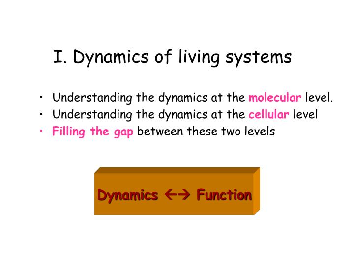
I. Dynamics of living systems. Understanding the dynamics at the molecular level. Understanding the dynamics at the cellular level Filling the gap between these two levels. Dynamics Function. Life’s complexity pyramid. Oltvai & Barabasi, Science 2002, 298, 763-764.

E N D
I. Dynamics of living systems • Understanding the dynamics at the molecular level. • Understanding the dynamics at the cellular level • Filling the gap between these two levels Dynamics Function
Life’s complexity pyramid Oltvai & Barabasi, Science 2002, 298, 763-764
“The complexity pyramid might not be specific only to cells” Different levels of structural organization: Dominance of molecular machinery Increasing specificity/chemistry) microtubules
Challenge:to understand thelong-time dynamics of large systems Model: Coarse-grained Method:Analysis of principal modes of motion (Frame transformation:Cartesian collective coordinates)
What is the optimal (realistic, but computationally efficient) model for a given scale (length and time) of representation? • Which level of details is needed for representing global (collective) motions? • How much specificity we need for modeling large scale systems and/or motions? • What should be the minimal ingredients of a simplified (reductionist) model?
Protein dynamics • Folding/unfolding dynamics • Passage over one or more energy barriers • Transitions between infinitely many conformations • Fluctuations near the folded state • Local conformational changes • Fluctuations near a global minimum
Can we predict fluctuations dynamics from native state topology only? Gaussian Network Model FOR MORE INFO… Bahar, I., Atilgan, A.R., & Erman, B. (1997) Folding & Des. 2, 173. Flory, P.J. (1976) Proc. Roy. Soc. London A. 351, 351.
Detailed specific potentials Approximate uniform potential “A single parameter potential is sufficient to reproduce the slow dynamics in good detail”
Rouse chain Connectivity matrix • Vtot = (g/2) [ (DR12)2 + (DR23)2 + ........ (DRN-1,N)2 ] • = (g/2) [ (DR1 -DR2)2 + (DR2 -DR3)2 + ........
Vtot = (g/2) DRT GDR Kirchhoff matrix of contacts 1 if rik < rcut 0 if rik > rcut Gik= G = Gii = -Sk Gik
Comparison with X-ray Temperature Factors 3 Bahar, I., Atilgan, A.R., & Erman, B. (1997) Folding & Design 2, 173-181 Debye-Waller factors: Bk = 8 2 <Rk Rk> /3 FOR MORE INFO...
Comparison with H/D Exchange – NMR data DSi = k ln W(DRi) = - g (DRi)2/ (2T [G-1]ii) Bahar, I., Wallquist, A., Covell, D.G., and Jernigan, R.L. (1998) Biochemistry 37, 1067.
Covariance matrix(directly found from MD or MC trajectories) C = DRi = instantaneous fluctuation in the position vector Riof atom i= Ri - <Ri> <DR1 . DR1> = ms fluctuation of site 1 averaged over all snapshots.
Eigenvalue decomposition of C Time-consuming aspect of PCA U is the matrix of eigenvectors,L is the diagonal matrix of eigenvalues. The ith column (eigenvector) of U is given by a linear combination of Cartesian coordinates and represents the axis of the ith collective coordinate (principal axis) in the conformational space. C = U L U-1 The ith eigenvalue represents the mean-square fluctuation along the ith principal axis. The motion along the ith principal axis is the ith mode.
Decomposition into normal modes • Slowest (global) modes function • Fastest (local) modes stability FOR MORE INFO... Bahar, I., Atilgan, AR, Demirel MC, Erman B. (1998) Physical Review Lett. 80, 2733. Demirel MC, Atilgan AR, Jernigan RL, Erman B. & Bahar, I. (1999) Protein Science 7, 2522.
Compare experimental B-factors with theoretical B-factors http://www.ccbb.pitt.edu/CCBBResearchDynHemRel.htm
T 92-100 2 35-40 145-146 b2 37-44 84-94 1 132-141 Comparison of the slowest modes of T and R2 b chain a chain R2 0 50 100 150 200 250 300 residue number
T R transitions in Hb Experimental T Experimental R2 Computed (R2) Reference... Xu & Bahar, submitted.
Order parameters for CO-bound and unliganded Hb Si = 3/2 <cos2Dai> - 1/2 For details on theory see... Haliloglu & Bahar, Proteins 1999, 37, 654-667
(I) (II) (A) (B) (I) (II) Fluctuations of the nevirapine-bound (A) and unliganded (B) forms of RT • Fluctuating conformations of the nevirapine-bound (A) and unliganded (B) forms of RT. The p66 subdomains are colored cyan (fingers), yellow (palm), red (thumb), green (connection) and pink (RNase H). • See the difference in the mechanism of global fluctuations for the liganded and unliganded RTs. This difference is significant given that the sizes or distributions of fluctuations are unaffected by ligand binding http://www.ccbb.pitt.edu/CCBBResearchDomMot.htm
Two hinge bending sites • (A) Two hinge-bending centers on RT forming minima in Figure 1: (I) near the NNRTI binding site, involving residues 107-110 (cyan),161-165 (green),180-188 (red) and 219-231 (blue), and (II) near the p66 connection and RNase H interface, comprising residues 363-366 (cyan),394-408 (green),410-423 (loop, magenta),424-429 (interdomain linker, red), and 504-512 (yellow). • (B) A closer view of region II, showing explicitly the side chains near the hinge site. Close tertiary contacts are indicated by the yellow dots.
Topology-based models • Folding/unfolding processes (folding loss of configurational entropy) • Micheletti et al, PRL (1999) • Cecconi et al. Proteins (2001) • Go & Scheraga Macromolecules (1976) • Galzitskaya & Finkelstein, PNAS (1999) • Munoz et al. PNAS (1999) • Alm & Baker, PNAS (1999) • Klimov & Thirumalai, PNAS (2000) • Clementi et al (Onuchic), JMB (2000) • Near-native fluctuations (springs acting on effective centroids, usually Ca atoms) • Ben-Avraham (1993) • Tirion (1996) • Bahar et al. (1997) • Hinsen (1998) • Sanejouand, Tama (2000) • Wriggers, Brooks (2001) • Ma (2002) “Native topology determines force-induced unfolding pathways”
Protein folding kinetics examined by a Go-like model Koga, N. & Takada, S. J Mol. Biol. 2001, 313, 171-180
Topological and Energetic Factors: What determines the transition state ensemble, and folding intermediates? Simulations with Go-like potential Applied to CI2, SH3 (2-state folders) and barnase, RNase H and CheY (have intermediates) “ Topology plays a central role in determining folding mechanisms” Can we use such simplified approaches for estimating amyloidogenic intermediates? Clementi, C. Nyemeyer, H. & Onuchic, J. N. J Mol. Biol 2000, 298, 937.