Download
1 / 45
450 likes | 661 Views
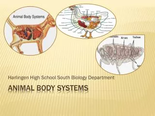
Animal Systems . Organization. All living things made up of cells Groups of cells work together to form tissues Groups of tissues organs organ systems There are 4 basic tissue types in animals: Epithelial: covering ( eg skin) keeps germs out and protects the body and organs

E N D
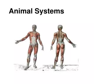
Animal Systems
Organization • All living things made up of cells • Groups of cells work together to form tissues • Groups of tissues organs organ systems • There are 4 basic tissue types in animals: • Epithelial: covering (eg skin) keeps germs out and protects the body and organs • Muscle: can contract-relax producing movement • Connective: supports and holds body together (tendons, ligaments, bone, cartilage)...also blood • Nerve: generates electrical signals for communication within the body
Circulatory System • Transportation of nutrients, gases and wastes • Defense against infection
Digestive System physical and chemical breakdown of food absorption of nutrients
Respiratory System gas exchange O2 in and CO2 out
Homeostasis The body is always trying to maintain a steady state or balance there are normal values or ranges: ·temperature 36.2-37.2oC ·heart rate 50-100 beats/min ·breathing rate 16-20 breaths/min ·blood pressure 120/80 mmHg homeostasis is maintained by feedback systems
Circulatory System
Main function: pumping blood throughout the body for: 1. Transport of nutrients from digested food (digestive) 2. Transport of oxygen from the lungs and carbondioxide to the lungs (respiratory) 3. White Blood cells fight off infection 4. Control of body temperature
There are 3 basic cycles for blood flow: • Cardiac circulation – blood capillaries within the heart • Pulmonary circulation – from the heart to the lungs • Systemic circulation – from heart to the rest of the body
Closed Circulatory system • All blood is contained within the blood vessels and the heart. • Deoxygenated blood to the RA • RV pumps deoxygenated blood to the lungs • Oxygenated blood to the LA • LV pumps oxygenated blood to the rest of the body
Blood Vessels • Blood vessels carry the blood around the body. • Arteriescarry blood away from the heart • thick walls of smooth muscle and elastic tissue to allow for expansion with the pressure • usually carry oxygenated blood to tissues • Veinscarry blood to the heart • one way valves prevent backflow of blood • weakened valves result in varicose veins. • usually carry deoxygenated blood to heart • Capillariesare tiny vessels, one cell thick connect the veins and arteries.
Blood • 55% of volume is plasma (mostly water) • carries glucose, vitamins, minerals, blood proteins, waste products and dissolved gases • Red Blood Cell (Erythrocytes) • RBC= 40% of the blood volume • contain millions of haemoglobin molecules...oxygen binds to iron • carry O2 from the lungs to the body tissues • carry CO2 from the body tissues back to the lungs
White Blood Cells (Leukocytes) • protect the body from infections • five types of white blood cells based on nucleus shape • Blood Platelets • cell-like fragments, smaller than the other blood cells • help in the blood clotting…clump together • Fibrin, a chemical is released to aid in the clotting process
Learning Check Q#4,5,6,7,8,10,11 Chapter 12.1 Review
Heart Beat "lubb-dubb" sound. • Heart “relaxed”…LA and RA fill with blood...diastole. • atria contracts and blood moves into ventricles • ventricles contract pushing the blood out of the heart...the blood is put under pressure...systole. • closing valves...atria to the ventricles=“lubb” • closing valves...ventricles to arteries= “dubb”
Blood Pressure • pressure against the arterial walls…measured with a sphygmomanometer. • pressure when the heart contracts (systolic) • and relaxes (diastolic). • Blood pressure= systolic pressure diastolic pressure
Control of Heart Rate • contraction without brain input • Sinoatrial(SA) node, also called pacemaker cells are nerve cells in the right atrium • SA node causes the atria to contract • sends electrical stimuli to the atrioventricular (AV) node. • electrical stimuli are sent through two nerve fibres called Purkinje fibres to the ventricles • causing them to contract.
Comparative Anatomy • Open Circulatory System– • Blood is pumped into the body cavity where tissues “bathe”. • oxygen diffuses into the tissues • Small organisms • Other Closed circulatory systems: • Amphibian– • three chambered heart. • blood mixed in ventricle • Efficient??
Fish– two chambered heart. • Deoxygenated blood enters atrium, then into the ventricle • pumped to the gills for oxygenation • blood then flows throughout the body…returning to the heart
Circulatory Disorders page 494 1. What is arteriosclerosis? 2. Describe the two methods of treating arteriosclerosis 3. What is an aneurysm? 4. What is an arrhythmia? 5. What are congenital defects? Describe heart murmurs as an example 6. What is a stroke? 7. Describe 3 technologies used in diagnosing: ECHO, ECG, Holter monitor 8. Answer page 498#19-22, 24 9. What are the following blood disorders: hemophilia and leukemia?
Circulatory Disorders page 494 1. What is arteriosclerosis? ...arteries thicken and lose elasticity 2. Describe the two methods of treating arteriosclerosis ...angioplasty-insert a tube into artery, and a tiny balloon forces the artery open ...coronary bypass 3. What is an aneurysm?...bulge in the artery caused by weakened area 4. What is an arrhythmia?...irregular heartbeat 5. What are congenital defects? Describe heart murmurs as an example....born with....heart murmurs abnormal blood flow in the heart (example valves do not close fully causing a wooshing sound as blood leaks through when the heart beats) 6. What is a stroke?...arteries supplying blood to the brain are damaged results in brain tissue not getting nutrients and oxygen 7. Describe 3 technologies used in diagnosing: ECHO, ECG, Holter monitor ...ECHO (echocariogram)- ultrasound to see 3D of heart...could check valves and chambers ...ECG (electrocardiogram) ...Holter monitor records heart signals for 24-48 hours 9. What are the following blood disorders: hemophilia nad leukemia?
12.1 review page 488#2,4,7,9 12.2 review page 493#1,2,6,7,9 Circulatory Review page 513#1-3,6,7,9,14,15,26,27,35 ,
Respiratory System
Respiration includes: 1. Ventilation:inhalation of O2 and exhalation of CO2 2. External Respiration: exchange of gases between alveoli (lungs) and blood (circulatory) 3.Internal Respiration: movement of gases between the blood and the cells
Nasal cavity: (moistens, warms, cleans and filters (cilia)the air) • Oral cavity: cleaning and warming steps missed • Pharynx: path for air and food • Epiglottis: directs food to the esophagus • Larynx: cartilage (vocal chords)..air passes=sound • Trachea: the windpipe, C- shaped cartilage keeps it open • Bronchi: 2 cartilage branches. Cilia and mucus prevents foreign materials (dust) from going on • Lungspecialized organ for gas exchange • bronchioles...Alveoli...gas exchange with capillaries • Ribs: protection and structural support
Mechanisms of Breathing (Ventilation) • 1.Inhalation: • Diaphragm (thin muscle-bottom of thoracic cavity) contracts and moves down • Intercostal muscles contract...ribs move up and out • Increases the thoracic cavity (chest)volume which decreases air pressure • Air moves from outside (high pressure) into the lungs (low pressure) • 2. Exhalation: • Opposite of inhalation…Decreases the thoracic cavity volume…increases the pressure…Air moves out
Gas Exchange • Diffusion: random movement of molecules...high concentration to an area of low concentration • Alveoli are the grapelike structures at the ends of the bronchioles...network of capillaries • Capillary and alveoli membranes are one cell thick • Moist surface allows for diffusion of both CO2 and O2
Control of Breathing • Controlled by automatic (autonomic) nervous system • Medulla oblongata located at the stem of the brain • chemoreceptors detect changes in pH • • increase in blood CO2increase in carbonic acid (lower pH) increases breathing rate
Control of Breathing • How Puffers Restore Homeostasis: • Asthma attack – decrease in airway diameter (bronchoconstriction) – • Steroid puffer – bronchodilation – increases air flow • Anaphylactic shock – smooth muscles in the bronchioles swell and pinch the tube • Epipen– epinephrine causes the smooth muscles to relax
Respiratory Disorders: • Pneumonia: inflammation in one or both lungs; it is usually caused by a viral infection or a bacterial infection... interferes with gas exchange, and the body becomes starved for oxygen. • Bronchitis: bronchi become red, inflamed, and filled with mucus...acute (due to infection) or chronic (due to an irritant...cilia lining the bronchi are gradually destroyed) • Asthma:Inhaledirritants such as pollen, dust, and smoke can often trigger an inflammation of the bronchi and bronchioles...narrows the air passages of the bronchi and bronchioles, thus reducing airflow. People with asthma experience wheezing, coughing, tightness in the chest, and shortness of breath • Emphysema: walls of the alveoli lose their elasticity...the respiratory surface for gas exchange and causes an oxygen shortage...Exhaling becomes difficult because the small airways collapse during exhalation, trapping air in the lungs and blocking the airflow • Cystic Fibrosis: genetic condition causes cells lining the airways to release thick, sticky mucus that clogs the lungs, leading to difficulty in breathing. The mucus traps disease-causing agents, making it difficult to clear bacteria that cause lung infections
Review OERB Flash Quizzes Resp and Circ