Download
1 / 29
290 likes | 295 Views
Microstructure and mechanical properties of Zircaloy-4 claddings hydrogenated at temperatures typical for LOCA conditions. A . Pshenichnikov, J. Stuckert. QWS19, Karlsruhe 2013. Objectives. Structure assessment of annealed and hydrogenated specimens Fracture surface investigation

E N D
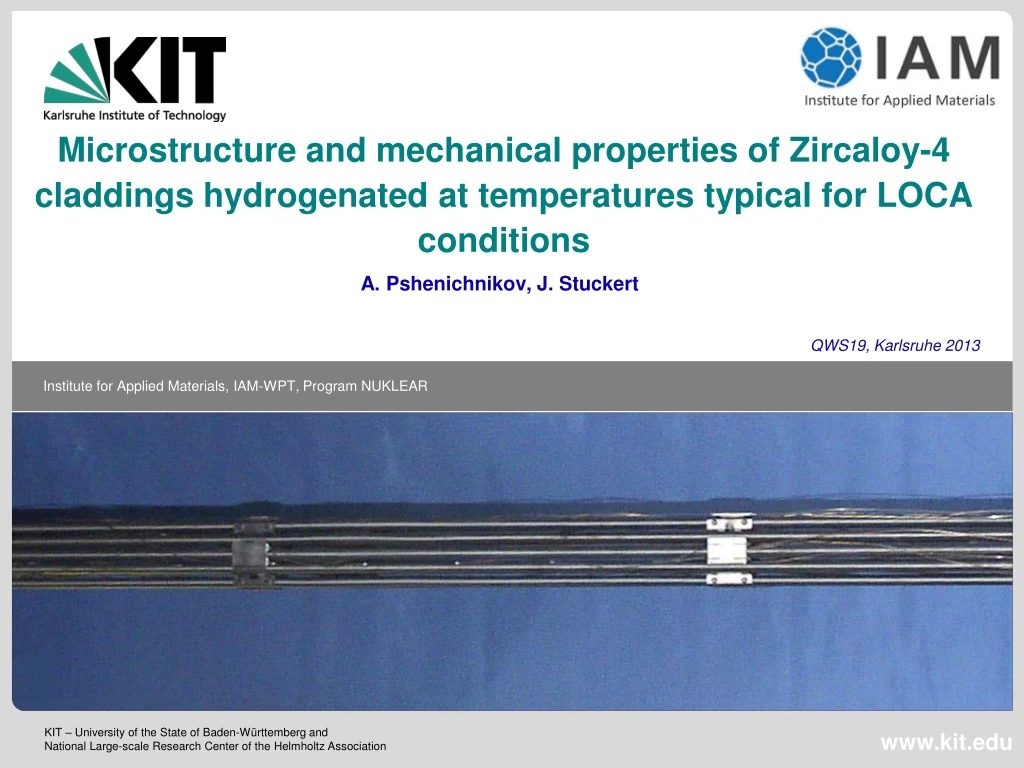
Microstructure and mechanical properties of Zircaloy-4 claddings hydrogenated at temperatures typical for LOCA conditions A. Pshenichnikov, J. Stuckert QWS19, Karlsruhe 2013
Objectives • Structure assessment of annealed and hydrogenated specimens • Fracture surface investigation • Zirconium hydrides detection • Progress in understanding the mechanism of embrittlement of Zirconium alloys • Application to the results of QUENCH-LOCA test
Equilibrium phase diagram of Zr-H system* • selected examples of our tests • Region of interest is LOCA conditions • Cooling in air • 700 wppm H • 9500 wppm H • * According to E. Zuzek et al., Bull. Alloy Phase Diagr. (1990), 385
Material and methods of investigation • Material: Conventional Zircaloy-4 cladding tube • ICP-OES measurement of Zircaloy-4 chemical composition (by weight):Sn: 1.33±0.02%, Fe: 0.23±0.002%, Cr: 0.12±0.0003%, O: 0.116±0.003%, Zr balance • Methods of investigation: • Hydrogenation in Ar+H2 gas mixture in LORA-furnace • Metallographic investigations and microhardness tests of the tube section • X-Ray diffraction analysis in the cladding tube wall middle • Scanning electron microscopy of polished and etched as well as fractured surfaces
Experimental procedure • Hydrogenation facilityLORA furnace • Specimen before hydrogenation • Specimen withdrawal in air after hydrogenation • Cooled specimen after hydrogenation • H2 duration was 2 to 12 minutes • Hydrogen gas partial pressure was 0.1 bar • Estimated cooling rate was 5 K/s
Optical metallography of annealed Zircaloy-4 • Annealing of Zircaloy-4 cladding at various temperatures during 8 minutes and fast cooling in air As-received 700 °C At 810 °C starts α→βphase transformation 900 °C 800 °C
Optical metallography of hydrogenated Zircaloy-4 • Hydrogenation of Zircaloy-4 cladding at 600 °C in Ar+H2 mixture and fast cooling in air 780 wppm H 1400 wppm H 2000 wppm H 3490 wppm H
Optical metallography of hydrogenated Zircaloy-4 • Hydrogenation of Zircaloy-4 cladding at 700 °Cin Ar+H2 mixture and fast cooling in air 1330 wppm H 2250 wppm H • α - Zr„islands“ 4000 wppm H 4760 wppm H
Optical metallography of hydrogenated Zircaloy-4 • Hydrogenation of Zircaloy-4 cladding at 800 °Cin Ar+H2 mixture and fast cooling in air 1790 wppm H 3060 wppm H 8600 wppm H 9550 wppm H
Optical metallography of hydrogenated Zircaloy-4 • Hydrogenation of Zircaloy-4 cladding at 900 °Cin Ar+H2 mixture and fast cooling in air 880 wppm H 1500 wppm H 2000 wppm H 6700 wppm H
Scanning electron microscopy of annealed and hydrogenated Zircaloy-4 After polishing and etching 0 wppm H 8600 wppm H
Scanning electron microscopy of annealed and hydrogenated Zircaloy-4 After polishing and etching 0 wppm H 8600 wppm H
Scanning electron microscopy of annealed and hydrogenated Zircaloy-4 After polishing and etching 0 wppm H α-Zr(O)? α-Zr (priorβphase)? 8600 wppm H γ- Zrhydrides? δ- Zrhydrides?
Scanning electron microscopy of fracture surfaces of hydrogenated Zircaloy-4 700 °C 720 wppm H 1860 wppm H 4850 wppm H 2790 wppm H
Scanning electron microscopy of fracture surfaces of hydrogenated Zircaloy-4 800 °C 1110 wppm H 3070 wppm H
Scanning electron microscopy of fracture surfaces of hydrogenated Zircaloy-4 900 °C 1170 wppm H 1640 wppm H Ductile fracture on needles of α-Zr(O) Brittle fracture
Microhardness of annealed and hydrogenated Zircaloy-4 hydrogen + T impactat 900 °C hydrogen impactat 600 °C • The diagonal length of resulting indented square was about 10 µm.
X-Ray profiles of Zircaloy-4 samples hydrogenated at 600 °C Hydridesare compressed with 0.5% of local strain
X-Ray profiles of Zircaloy-4 samples hydrogenated at 700 °C
X-Ray profiles of Zircaloy-4 samples hydrogenated at 800 °C
Change of lattice parameters after hydrogenation of Zircaloy-4 samples a δ – hydride ZrH1,66scheme γ – hydrideZrHscheme c a c 3D model of HCP-lattice Scheme of decohesion mechanism, accompanied by hydride formation 2D projection of HCP-lattice without hydrogen With hydrogen
Conclusion • No “macroscopic” hydrides were detected by means of optical microscopy, only high magnification SEM observations reveal presumable submicroscalehydrides. • Microhardness tests showed a relationship between hydrogen content hardening, annealing softening and hardening due to β → αtransformation during cooling phase. • The XRD-analysis showed the presence of γ-, δ-phases of zirconium hydrides in all of performed experiments. With the increase of hydrogen content the hydride peak intensity was also increased. Simultaneously the hydrogen should be partially dissolved in the lattice which is indicated by increase of the lattice parameter “c”. • Because carried out observations have proved only the presence of hydrides and gave not enough information on the hydride structure and distribution, further detailed EBSD and TEM investigations should be performed in order to determine the location, morphology and orientation of nano-scaled hydrides and to separate hydrides from other structural features. • The performed experiments can explain the absence of macroscopic hydrides in QUENCH-LOCA experiments. The increased brittleness of some zirconium claddings after QUENCH-LOCA tests could be caused by nano-scaled hydrides which are distributed in the bulk of material. The fact of the growth of the lattice parameter “c” allows to suggest that the decohesion mechanism accompanied by hydride formation could be responsible for cladding destruction.
Aknowledgements • The authors would like to thank the following KIT colleagues: • Dr. Mario Walter for fruitful discussions • Mrs. Ursula Peters for her technical assistance with the hydrogenation tests • Ms. Julia Lorenz for the help during preparation of the specimens for metallographic observations • Dr. HaraldLeiste for carrying out of X-Ray diffraction measurements • Dr. Marcus Müller for carrying out of FEG-SEM observations
Thank you for your attention! anton.pshenichnikov@kit.edu juri.stuckert@kit.edu http://www.iam.kit.edu/wpt/471.php