Download
1 / 24
240 likes | 252 Views
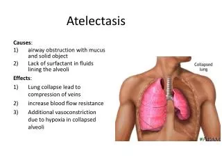
Chapter 42 Postoperative Atelectasis. B. A. B. A. Figure 42-1. Alveoli in postoperative atelectasis. A, Total alveolar collapse. B, Partial alveolar collapse. Anatomic Alterations of the Lungs. Alveoli of primary lobules (microatelectasis or subsegmental atelectasis)—very common

E N D
B A B A Figure 42-1. Alveoli in postoperative atelectasis. A, Total alveolar collapse. B, Partial alveolar collapse.
Anatomic Alterations of the Lungs • Alveoli of primary lobules (microatelectasis or subsegmental atelectasis)—very common • Lung segment—fairly common • Lung lobe—less common • Entire lung—rare
Etiology Decreased Lung Expansion • Good lung expansion depends on the patient’s intact chest cage and his or her ability to generate an appropriate negative intrapleural pressure. • Thoracic and upper abdominal procedures often result in a reduction in the patient’s ability to generate good lung expansion • And, therefore, are considered as high-risk factors for subsequent development of postoperative atelectasis.
Etiology (Cont’d) Decreased Lung Expansion • Other precipitating factors • Anesthesia • Postoperative pain • Supine position • Obesity • Advanced age • Inadequate tidal volumes during mechanical ventilation • Malnutrition • Ascites • Diaphragmatic apraxia • The presence of a restrictive lung disorders
Etiology (Cont’d) Alveolar Degassing • Postoperative atelectasis often is associated with • Retained airway secretions • Mucous plugs
Etiology (Cont’d) Alveolar Degassing • Precipitating factors include: • Decreased mucociliary transport • Excessive secretions • Inadequate hydration • Weak or absent cough • General anesthesia • Smoking history • Gastric aspiration • Certain preexisting conditions (e.g., chronic bronchitis, asthma)
Overview of the Cardiopulmonary Clinical Manifestations Associated with Postoperative Atelectasis The following clinical manifestations result from the pathophysiologic mechanisms caused (or activated) by Atelectasis
Clinical Data Obtained from Laboratory Tests and Special Procedures
PaO2 and PaCO2 trends during acute alveolar hyperventilation.
PaO2 and PaCO2 trends during acute or chronic ventilatory failure.
Figure 42-2 A, Endotracheal tube tip misplaced in the right main stem bronchus (arrow). Note that the left lung has collapsed completely (i.e., white fluffy appearance in the left lung). B, The same patient 20 minutes after the endotracheal tube was pulled back above the carina (arrow). Note that the left lung is better ventilated (i.e., appears darker).
General Management of Postoperative Atelectasis • Precipitating factors for postoperative atelectasis should be identified • High-risk patients should be monitored closely • Preventive measures should be prescribed for high-risk patients • Incentive spirometry • Chest physical therapy • Whenever possible, treatment of the underlying cause of atelectasis should be prescribed immediately
Respiratory Care Treatment Protocols Oxygen Therapy Protocol Bronchopulmonary Hygiene Therapy Protocol Lung Expansion Therapy Protocol Mechanical Ventilation Protocol