Download
1 / 30
300 likes | 515 Views
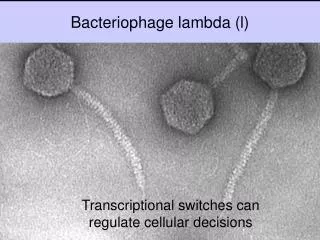
Interactions in macromolecular assemblies. Journey of bacteriophage M13 major coat protein. David Stopar. University of Ljubljana Slovenia. Journey of bacteriophage M13 major coat protein. Replication cycle of the bacteriophage M13 Structure of the major coat protein

E N D
Interactions in macromolecular assemblies Journey of bacteriophage M13 major coat protein David Stopar University of Ljubljana Slovenia
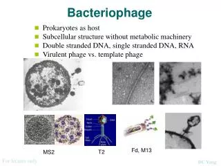
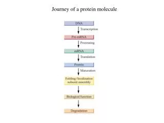
Journey of bacteriophage M13 major coat protein • Replication cycle of the bacteriophage M13 • Structure of the major coat protein • Topology of the major coat protein in the lipid bilayer • Anchoring of the major coat protein in the lipid bilayer
gp 8 M13 DNA gp 5 Schematic representation of bacteriophage M13 replication cycle E.coli F-pilus
Gene 7 protein Distal end Gene 9 protein Circular ssDNA Gene 8 protein Gene 6 protein Proximal end Gene 3 protein Schematic structure of bacteriophage M13 filament
Nearest-neighbour interactions between protein subunits in the phage particle k=11 k=11 k=6 k=6 k=0 k=0
Journey of bacteriophage M13 major coat protein • Replication cycle of the bacteriophage M13 • Structure of the major coat protein • Topology of the major coat protein in the lipid bilayer • Anchoring of the major coat protein in the lipid bilayer
Amino acid residue number 1 50 Major coat protein structural domains in lipid bilayer 1-5 N-terminus 6-16 amphiphatic helix 17-23 loop 24-46 transmembrane helix 47-50 C-terminus
X-ray and NMR limitations for membrane proteins • X-ray: “no” crystals of membrane proteins • NMR: membranes are anisotropic systems • high-resolution NMR limited to micellar systems • solid-state NMR is needed for bilayer structure • 13C and 15N-enrichment is needed (difficult and very expensive!)
? ? ? ? ? ? ? ? Structure not possible in a membrane M13 coat protein in SDS micelles 2D NMR & DG analysis Papavoine et al. (1998)J. Mol. Biol. 282, 401-419
Molecular Dynamics (MD) Approach Starting conformation for protein in POPC membrane from SDS (Papavoine et al., 1998).
0.5 ns 0 ns 1 ns 1.5 ns 42 H-bonds 41 H-bonds 43 H-bonds 41 H-bonds Snap Shots of Protein Backbone The effects seen in MD are in the ESR time scale (ns)
M13 Coat Protein with Labels Attached Note: one label is measured at a time membrane-water interface
Position withstructural restriction O • O N N O ESR spin label: 5-maleimido-proxyl Position with nostructural restriction ESR Spin Label Information
Amino acid residue number 1 50 2Azz Outer Splitting Sensitivity of spin labels for different protein sites
Journey of bacteriophage M13 major coat protein • Replication cycle of the bacteriophage M13 • Structure of the major coat protein • Topology of the major coat protein in the lipid bilayer • Anchoring of the major coat protein in the lipid bilayer
Amino acid residue number A49C 1 V31C G38C 50 Spin label relaxation times in DOPC bilayers
Amino acid residue number oxygen 1 Ni2+ 50 Relative quenching efficiency by oxygen and Ni2+ Ni2+
Amino acid residue number oxygen 1 Ni2+ 50 Dependence of spin labeled DOPC acyl chains on the relaxation enhancement by oxygen and Ni2+
Amino acid residue number 1 50 Location of the protein in the lipid bilayer as determined by fluorescence labeling
Topology of the major coat protein in the lipid bilayer Interface Leu 14 Trp 26 Hydrocarboncore Lys 40 Phe 45 NH3+ Interface 20 Å
Journey of bacteriophage M13 major coat protein • Replication cycle of the bacteriophage M13 • Structure of the major coat protein • Topology of the major coat protein in the lipid bilayer • Anchoring of the major coat protein in the lipid bilayer
Amino acid residue number 1 50 AEDANS lmax in DOPC lipid bilayers
Amino acid residue number 1 50 Tryptophane lmax in DOPC lipid bilayers
Phe 11 Leu 14 Lys 8 Trp 26 Lys 40 Lys 43 Lys 44 Phe 42 Phe 45 Bacteriophage M13 major coat protein anchors in the lipid bilayer N-terminus C-terminus
gp 8 M13 DNA gp 5 Schematic representation of bacteriophage M13 replication cycle E.coli F-pilus
(1) (2) (3) N-terminus C-terminus Model of the major coat protein disassembly and assembly
Acknowledgements Wageningen Marcus Hemminga Rob Koehorst Ruud Spruijt Werner Vos Cor Wolfs Ljubljana Ivan Mahne Janez Štrancar Milan Schara Göttingen Derek Marsh Kity A. Jansen Szeged Tibor Páli