Download
1 / 38
380 likes | 1.02k Views
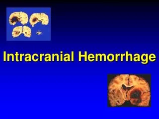
Intracranial hemorrhage. 영상의학과 강병관. 개요. 두개내 출혈은 매우 다양한 원인에 의해 발생하여 출혈의 원인이나 그 부위에 따라 다양한 임상적 의미를 갖는다 . 출혈의 소견을 이해하기 위해서는 출혈이 발생하는 배경 및 발생한 후 일어나는 변화에 대하여 이해할 필요가 있다 . 출혈의 방사선학적 소견에 영향을 미치는 인자. 1. 출혈의 원인 2. 출혈의 위치 및 정도 3. 출혈 후 시간경과 4. 환자의 혈액 및 응고기전의 상태 영상 방법 및 기법.

E N D
Intracranial hemorrhage 영상의학과 강병관
개요 • 두개내 출혈은 매우 다양한 원인에 의해 발생하여 출혈의 원인이나 그 부위에 따라 다양한 임상적 의미를 갖는다. 출혈의 소견을 이해하기 위해서는 출혈이 발생하는 배경 및 발생한 후 일어나는 변화에 대하여 이해할 필요가 있다.
출혈의 방사선학적 소견에영향을 미치는 인자 • 1.출혈의 원인 • 2.출혈의 위치 및 정도 • 3.출혈 후 시간경과 • 4.환자의 혈액 및 응고기전의 상태 • 영상 방법 및 기법
두개내 출혈의 다양한 원인 • Cranial Contusion - Epidural hematoma (EDH) - Subdural hematoma (SDH) - Subarchnoid hemorrhage (SAH) • Hypertension - Intracerebral hemorrhage (ICH)
Aneurysm 파열 - subarchnoid hemorrhage(SAH) • Anteriovenous malformation (AVM) 등 혈관성 질환 - ICH , SAH • 원발성 혹은 전이성 종양 • 뇌혈관 경색
고혈압성 출혈의 흔한 위치 및 발생빈도 • Putamen/external capsule 60~65% • Thalamus 15~25% • Pons 5~10% • Cerebellum 2~ 5% • Subcortical white matter 1~ 2%
출혈후 변화 • 혈액성분의 화학적 변화 -hemoglobin의 denaturation -blood degradation product
혈종의 물리적 변화 -martrix formation -clot retraction -병소부위 및 주위의 세포 침윤 -fibrinolysis
출혈의 시간경과에 따른병리학적 단계 (6단계) • Hyperacute (출혈후 6시간) -oxyhemoglobin (Ohb) -혈소판, 적혈구응집 -적혈구의 변화 discoid, biconcave -> spherical -fibrin mass 형성 -단백질 농도 증가 -병변주위 부종시작
Acute (7시간-3일) -부종뚜렷 -적혈구의 echinocyte -fibrin mass -> fibrinolysis -intracellular deoxyhemoglobin (DHb) -macrophage의 침윤
Subacute (4일-10일) -병변부위 모세혈관증식 -perivascular의 염증반응 -macrophage의 침윤, edema최대 -deoxyhemoglobin: DHb --> oxidation - met hemogrobin:MHb
Early chronic(11일-6주) -edema 소실 - gliosis -macrophage ferritin hemosiderin
Late chronic (7주- 6개월) -식세포의 적혈구 포획은 끝나고 병변 수축 • Ancient (6개월 이상) -영구적 상처 -hemosiderin 침착에의한 갈색의 국소적 병변
출혈부위에 따른 소견 • Intracerebral hemorrhage(ICH) • Subarchnoid hemorrhage(SAH) • Epidural hematoma(EDH) • Subdural hematoma(SDH)
Intracerebral hemorrhage(ICH) • ICH의 원인 -Hypertension -AVM -angiographically occult vascular malformation -Moya-moya disease -hemorrhage conversion of cerebra infarction -venous infarction -헤르페스 뇌염 -원발성 뇌종양의 출혈, hemorrhagic metastasis -amyloid angiopathy
Subarchnoid hemorrhage(SAH) • 비외상성 SAH의 원인 -Ruptured intracranial aneurysm 70-80% -Ruptured intracranial AVM 5% -Idiopathic, cryptic vascular malformation 5-10% -ICH 에 동반되는 경우 10% -Etc 2%
Epidural hematoma(EDH) • 원인: 경막과 두개골 사이의 혈관파열 • 호발부위 -동맥: middle meningeal artey anterior falcial artey 추경동맥의 경막분지 -정맥: diploic vein meningeal emissary -정맥동: superior sagittal 정맥동 transnerser 정맥동
특징 -85% 정도의 동반골절 -주로 동맥파열에 의한 고압의 출혈 (혼수상태유발) • CT상의 모양 -동맥,정맥동 파열: 렌즈, 반달모양 -작은 정맥파열: 일부반달, 초생달 모양
지연성 경막외 혈종 (재출혈) -Slow venous oozing -수축되었던 손상혈관의 이완 -혈종내 모세혈관의 파열 -약물치료나 수술후 relief of tamponade effect
자연흡수 및 감소 -자연흡수 예방척도 없음 50cc이하 일때 자연흡수 경향이 높음 -빠른 크기 감소 골절 부위를 통한 EDH의 두개강외로의 이동
Subdural hematoma(SDH) • 원인: 피질과 정맥동 사이의 혈관파열 주- 피질교량 정맥 파열 이외- 정맥동 작은 피질동맥의 분지 • CT상의 모양 -주로 초생달 모양 -반달모양 -드물게 렌즈모양(만성 경막하 혈종)
CT상 동일밀도로 보이는 급성 경막하혈종 -헤모글로빈 농도 8~11gm% -혈장 알부민 농도 10~13gm% -만성 경막하 혈종내의 재출혈 -뇌척수액과 혼합 -MRI로 쉽게 진단.
경막하 혈종의 크기 증가 원인 -손상된 혈관의 재출혈 -혈종내 모세혈관의 파열 -혈종 삼투압증가 -약물치료나 수술후 Relief of tamponade effect
참고문헌 -신경방사선과학(서울대 의과대학) 장기현,김인원,한문희 공저 -일조각출판