Download
1 / 31
690 likes | 1.21k Views
Respiratory Physiology. Functions and organization of the respiratory system. Dr. Aida Korish Associate Prof. Physiology KSU. Learning Objectives. By the end of this lecture you will be able to:- 1- Describe the structures and functions of the conductive and respiratory zones of airways.

E N D
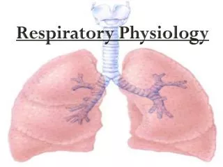
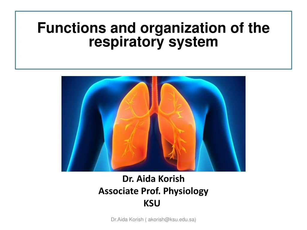
Respiratory Physiology Functions and organization of the respiratory system Dr. Aida Korish Associate Prof. Physiology KSU Dr.Aida Korish ( akorish@ksu.edu.sa)
Learning Objectives • By the end of this lecture you will be able to:- 1-Describe the structures and functions of the conductive and respiratory zones of airways. 2-Distinguish the difference between internal and external respiration. 3-Discuss the functions of the respiratory system, including non-respiratory functions, like clearance mechanism by mucus and cilia, production of surfactant and its physiological significance. Dr.AidaKorish ( akorish@ksu.edu.sa)
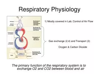
The main goal of respiration is to 1-Provide oxygen to tissues. 2- Remove CO2 from the body. Respiratory system consists of: • Passages (airways) • Muscles • Centers Dr.Aida Korish ( akorish@ksu.edu.sa)
Functions of the respiratory system include • Gas exchange (respiratory function). • Phonation: is the production of sounds by the movement of air through the vocal cords. • Pulmonary defense: the respiratory mucus membrane has muco-cilliary barrier filterand it produces • Immunoglobulin A (Ig A), • Alpha-1 antitrypsin, In addition, the pulmonary macrophages in the alveoli: engulf smaller foreign particles which pass through the muco-cilliary barrier filter. Dr.Aida Korish ( akorish@ksu.edu.sa)
Cont..non respiratory functions of lung • AngiotensinI is converted to angiotensin II with the help of angiotensin converting enzyme formed by the lungs. • Regulating the acid- base status of the body by washing out extra carbon dioxide from the blood. • Secretion of important substances like surfactant. Dr.Aida Korish ( akorish@ksu.edu.sa)
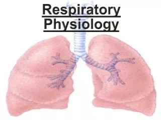
Respiratory passages (airways) Dr.Aida Korish ( akorish@ksu.edu.sa)
Organization of the respiratory system Dr.Aida Korish ( akorish@ksu.edu.sa)
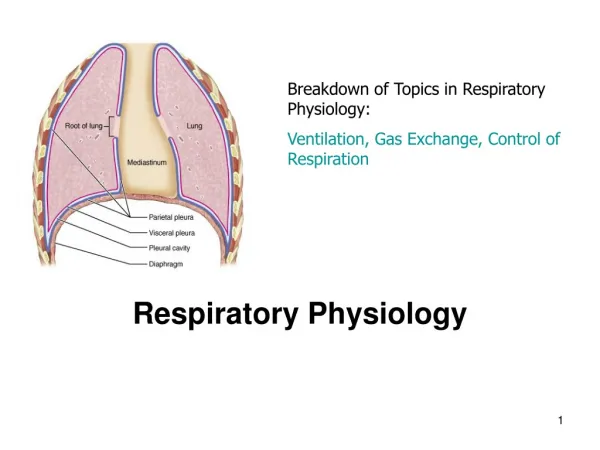
I- Conductive Zone II- Respiratory Zone (Respiratory unit) Includes: Respiratory bronchioles, alveolar ducts, alveolar sacs, alveoli Function in gas exchange. • Starts from nose to the end of terminal bronchioles. • Help warming, humidification, filtration of inspired air. • Contains the olfactory receptors for smell sensation. • Conducts the sound during speech. • Protective function by cough and sneezing reflexes. Dr.Aida Korish ( akorish@ksu.edu.sa)
External and Internal Respiration External respiration: is the process of gas exchange between the alveolar air and the pulmonary capillary blood. Internal Respiration: is the process of gas exchange between the blood in the systemic capillaries and the tissues. Dr.Aida Korish ( akorish@ksu.edu.sa)
External respiration 3 major functional events occurs during it: 1-Pulmonary ventilation: inward and outward movement of air between lung and atmosphere. 2- Diffusion of oxygen and CO2 between the alveoli and the pulmonary capillary blood. 3- Transportof O2 & Co2 in the blood and body fluids to and from the cells. Respiration (breathing) could be either: Resting breathing: normal breathing during resting conditions. Forced (maximal) breathing: It occur during exercise and in patients with bronchial asthma, allergy, other pulmonary diseases.
Lining cells of the alveoli 1- Type I alveolar epithelial cells ( type I pneumocytes) *Participate in the respiratory membrane , across which gas exchange takes place. 2- Type II alveolar epithelial cells ( type II pneumocytes) ( 10% of the surface area of alveoli) *Secrete surfactant. 3- Alveolar macrophages *Engulf the foreign bodies that reach the alveoli.
Surface Tension H2O molecules at the surface of alveoli are attracted to each other by attractive forces that resist distension called surface tension. • Surface tension tends to oppose alveoli expansion. • Pulmonary surfactant reduces the surface tension of the fluid lining the alveoli. • Collapsing Pressure is Caused by Surface Tension and is indirectly related to the size of alveoli ( law of LaPlace) . Dr.Aida Korish ( akorish@ksu.edu.sa)
Surfactant • Surfactant is a complex compound containing phospholipids esp. dipalmitoylphosphatidyl choline and a number of Apo proteins. • The earliest detection of surfactant from fetal alveoli begins between 6-7th month but this could be delayed in others to wk 35 of intrauterine life. • Surfactant reduces surface tension throughout the lung,reducing the effort required by the respiratory muscles to expand the lungs,prevents alveolar collapse, decreases airway resistance and decreases work of breathing.
Surfactant deficiency • Deficiency in premature babies causes respiratory distress syndrome of the new born (RDS) (hyaline membrane disease). • Prevention: Corticosteroid injection to mothers expected to deliver prematurely. This will enhance surfactant maturation. • After delivery they are given inhaled surfactant. • Smoking in adults, hypoxia or hypoxemia, decrease the secretion of surfactant and cause adult respiratory distress syndrome. Dr.Aida Korish ( akorish@ksu.edu.sa)
Innervations of lungs and bronchi • Is by autonomic nerves. • Sympathetic stimulation releases epinephrine (adrenaline) causes dilatation of the bronchi. • Parasympathetic stimulation releases acetyl choline causes constriction of the bronchi. • Locally secreted factors :histamine, slow reacting substances of anaphylaxis (SRSA) secreted by the mast cells due to allergy (as in patients with asthma) often cause bronchiolar constriction and increased airway resistance leading to forced breathing. Dr.Aida Korish ( akorish@ksu.edu.sa)
Mechanics of pulmonary ventilation Dr.Aida Korish ( akorish@ksu.edu.sa)
Learning Objectives • By the end of this lecture you will be able to: 1- List the muscles of respiration and describe their roles during inspiration and expiration. 2- Identify the importance of the following pressures in respiration: atmospheric, intra-alveolar, intrapleural, and transpulmonary. 3- Explain why intrapleural pressure is always subatmospheric under normal conditions, and the significance of the thin layer of the intrapleural fluid surrounding the lung. 4- Define lung compliance and list the determinants of compliance. Dr.Aida Korish ( akorish@ksu.edu.sa)
Role of muscles of respiration in ventilation Dr.Aida Korish ( akorish@ksu.edu.sa)
Cont…respiratory muscles Inspiratory muscles: - During resting inspiration are the diaphragm, external intercostal. - During forced inspiration the Accessory muscles of inspiration e.g sternomastoid, anterior serratus, scalene muscles contract in addition to the muscles of resting inspiration. Expiratory muscles: Resting expiration is a passive process that depends on the recoil tendency of the lung and need no muscle contraction. However, forced expiration is an active process and need contraction of 1-the Abdominal musclesand 2- the internal intercostal muscles. .
Deep Forceful Breathing • Deep Inspiration • During deep forceful inhalation accessory muscles of inspiration participate to increase size of the thoracic cavity • Sternocleidomastoid – elevate sternum • Scalene – elevate first two ribs. • Anterior serrati; elevates many of the ribs. • Pectoralis minor – elevate 3rd–5th ribs • Deep Expiration • Expiration during forceful breathing is an active process. • Muscles of exhalation increase pressure in abdomen and thorax • Abdominal muscles. • Internal intercostals.
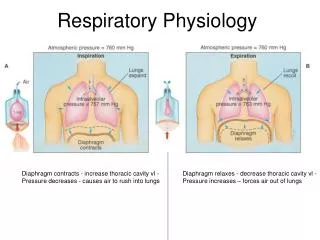
Pressure changes in the lungs during breathing Air will flow from a region of high pressure to one of low pressure-- the bigger the difference, the faster the flow
Intra-alveolar pressure(intrapulmonary pressure 1-Intra-alveolar Between breathes = zero pressure During inspiration = (-1 mmHg) and air (tidal volume) flows from outside to inside the lungs). At the end of inspiration = zero and air flow stops. During expiration = (+1 mmHg) and air flows out of the Lungs Dr.Aida Korish ( akorish@ksu.edu.sa)
2-Intrapleural pressure (IPP): Pressure in the pleural space is negative with respect to atmospheric pressure at the end of normal expiration (-5cmH2O). • Why negative??: 1- The lung's elastic tissue causes it to recoil, while that of the chest wall causes it to expand. Because of these two opposing forces the pressure in the pleural cavity becomes negative. 2-The pleural space is a potential space, (empty) due to continuous suction of fluids by lymphatic vessels. Dr.AidaKorish ( akorish@ksu.edu.sa)
Values of IPP • During resting position between breathes it = (-5) cm H2O. During resting inspiration it becomes more –ve (-7.5) cm H2O. • Forced ventilation Insp. :-20 to - 40 cm H2O Exp. : + 30 cm H2O Dr.Aida Korish ( akorish@ksu.edu.sa)
3-Transpulmonary pressure (TPp) (Extending Pressure) • The difference between the alveolar pressure (Palv) and the pleural pressure(Ppl). TPp = Palv-Ppl • It is a measure of the elastic forces in the lungs that tend to collapse the lungs (the recoil pressure). • It prevents lung collapse. • The bigger the volume of the lung the higher will be its tendency to recoil. Dr.AidaKorish ( akorish@ksu.edu.sa)
oCompliance of the lung (CL) The extent to which the lungs will expand for each unit increase in the transpulmonary pressure is called the lung compliance. CL = (∆ V) (∆ P) i.e the ratio of the change in the lung volume produced per unit change in the distending pressure. For both lungs in adult = 200 ml of air /cm H20. For lungs and thorax together = 110 ml/cm H20. Dr.Aida Korish ( akorish@ksu.edu.sa)
Compliance of the lungs • Compliance Diagram of the Lungs.. • The characteristics of the compliance diagram are determined by the elastic forces of the lungs. These can divided into (1) 1/3 is due to elastic forces of the lung tissue itself ( elastin, collagen). (2) 2/3 of the elastic forces caused by surface tension of the fluid that lines the inside walls of the alveoli and other lung air spaces. Dr.Aida Korish ( akorish@ksu.edu.sa)
Diseases that affect compliance of lung • Lung compliance is reduced in pulmonary fibrosis , pulmonary edema, diseases of the chest wall ( kyphosis, scoliosis, paralysis of the muscles, etc…). • Emphysema increases the compliance of the lungs because it destroys the alveolar septal tissue rich with elastic fibers that normally opposes lung expansion. Dr.Aida Korish ( akorish@ksu.edu.sa)