Download
1 / 56
570 likes | 616 Views
Common Eye Conditions and its defects. eyes.

E N D
eyes Your eyes are your body's most highly developed sensory organs. In fact, a far larger part of your brain is dedicated to the functions of eyesight than to those of hearing, taste, touch or smell. We tend to take eyesight for granted, yet when vision problems develop, most of us will do everything in our power to restore our eyesight to normal.
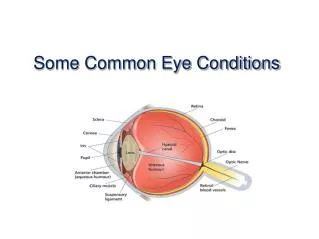
eye: camera The eye functions much like a camera. Light passes through the cornea and the lens of the eye and is focused on the retina in much the same way that an image is focused on the film of a camera.
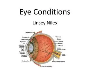
parts The eye is protected by 3 layers: sclera, choroid, and the retina. The sclera is the white covering of the eye. It is continuous with the cornea in the front. • The middle layer, the choroid, is a vascular layer lining the posterior (back) 3/5 of the eyeball. It is continuous with the ciliary body and the iris, which is the colored part of your eye. • The intermost layer is the retina, which is light-sensitive and has rods and cones.
parts you can see… The pupil is the small black opening in the iris. The iris controls the size of the pupil with 2 muscles: dilator muscle, which enlarges the pupil and the sphincter muscle, which makes the pupil smaller. The iris is the colored disk which is right behind the cornea. It's color comes from melanin. The more melanin there is and the closer it is to the surface, the darker the iris.
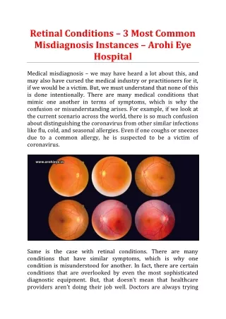
The retina Retinal blood vessels Macula (fovea in centre) Optic nerve
Red Eyes The "red eye" effect shown in this photograph is due to light reflection by the retina
visual acuity test Normal vision is measured at 20/20. This means that you can see clearly at 20 feet what should normally be seen at that distance. If you have 20/100 vision, that means that you must be as close as 20 feet to see what a person with normal vision can see at 100 feet. 20/20 vision is not perfect vision, it just indicates the visual acuity. There are many other things such as peripheral vision, eye coordination, depth perception, focusing ability and color vision that affect overall vision ability.
myopia and hyperopia • Nearsightedness and farsightedness have to do with the way the eye brings images into focus on the back of the eyeball, where 10 layers of light-sensitive nerve tissue make up the retina.
nearsightedness… Nearsightedness, or myopia, which affects about 20% of the population, is the result of images being focused in front of the retina rather than on it, so distant objects appear blurred. A nearsighted person holds a book close to the eyes when reading and has to sit in the front of the classroom or movie theater to see clearly. The condition runs in families and affects men and women equally, usually appearing in childhood and stabilizing in the twenties.
farsightedness… Farsightedness, or hyperopia, is the opposite of nearsightedness: The lens of the eye focuses images slightly behind the retina, making nearby objects appear blurry. Children often overcome mild farsightedness as they grow up and the eye muscles contract.
astigmatism… occurs when the eye lacks a single point of focus. The condition is a result of an uneven curvature of the cornea or, in some cases, an abnormality in the lens. People with astigmatism have a random, inconsistent vision pattern, in which some objects appear clear and others blurry.
test forastigmatism… An irregular curvature of the cornea and/or lens is determined using this type of illustration. When using the test card, a person would know if he has astigmatism becausesome radial lines would be blurred.
red-green colorblindness… is a disorder of the retina's light-sensitive cone cells, which respond to colors. Most people with color blindness see colors normally in bright light but have difficulty distinguishing reds and greens in dim light. Color blindness occurs mostly in men, afflicting 8% of the male population. It is extremely rare for someone to be totally color-blind --able to see only shades of gray.
Common eye conditions - prevalence 80 per cent of vision impairment and blindness in the population over the age of 40 is caused by five conditions (listed alphabetically): • Age-related Macular Degeneration (AMD) – 10 per cent • Cataract - 15 per cent • Diabetic retinopathy - 2 per cent • Glaucoma - 5 per cent • Under-corrected or uncorrected refractive error - 59 per cent
What is age-related macular degeneration (AMD)? • A chronic degenerative condition that affects the central vision. • progression of the condition is likely • ten per cent of people with macular degeneration have the “wet form” which may respond to treatment • the majority of people have the “dry form” • two out of three people will be affected by AMD in their lifetime.
Prevalence and risk factors of AMD • Ageing is the greatest risk factor with prevalence trebling with each decade over 40 years • AMD is present in 13 per cent of people between the ages of 70-75 and is the leading cause of vision impairment in Australia • Smoking increases the risk of developing AMD • Family history is also a risk factor - genes have been identified and linked with AMD
Functional implications of AMD • Difficulty distinguishing people's faces • Difficulty with close work • Perceiving straight lines as distorted or curved • Unable to differentiate between the footpath and road • Difficulty identifying the edge of steps if there is no colour contrast • Unable to determine traffic light changes • Difficulty reading, with blurred words and letters running together
Treatment of AMD • Treatment options are improving with new technology • The wet form can be treated with intravitreal injections that aim to prevent further vision loss • Lost vision cannot be recovered - early detection to identify those who can receive treatment is the key
Prevention of AMD • Early detection of AMD is crucial: • In the wet form of the disease, vision loss may be arrested with early treatment by an ophthalmologist • Regular eye examinations are the key to early detection of disease before vision loss occurs • If there are any changes in the quality of vision, refer to GP to arrange an appropriate referral to an eye health professional • Advise your clients to stop smoking
What is a cataract? • A cataract is the clouding of the lens inside the eye. With a cataract, light is scattered as it enters the eye, causing blurred vision
Prevalence and risk factors of cataract • 31 per cent of the population over the age of 55 has a cataract • Long term use of corticosteroids can increase risk of cataracts • Exposure to UV light can also increase the risk • Ageing, smoking and having diabetes can increase the risk of developing cataract.
Functional implications of cataract • Blurred vision • Reduced contrast • Having difficulty judging depth • Seeing a halo or double vision around lights at night • Seeing images as if through a veil/smoke • Being particularly sensitive to glare and light • Having dulled colour vision.
Treatment of cataract • Updating glasses can help with early cataract • Surgery: 180,000 cataract operations are done • usually in and out of hospital on same day • no general anaesthetic is required (in most cases) • the cloudy lens inside the eye is removed, except for the back capsule • an intraocular lens implant (IOL), a new lens is inserted into the eye
What is diabetic retinopathy? • This condition is a complication of diabetes • It affects the small blood vessels of the retina • Blood vessels begin to leak and bleed inside the eye
Prevalence and risk factors of diabetic retinopathy • It is estimated that three per cent of the population aged over 55 years have diabetic retinopathy • 22 per cent of people with known Type 2 diabetes have some form of retinopathy related to their diabetes • Within 15 years of being diagnosed with diabetes, three out of four diabetics will have diabetic retinopathy • People who have had diabetes for many years, have diabetic kidney disease or have Type 1 diabetes have a greater risk of developing diabetic retinopathy
Functional implications of diabetic retinopathy • Difficulty with fine details (e.g. when reading or watching television) • Fluctuations in vision from hour to hour or day to day • Blurred, hazy or double vision • Difficulty seeing at night or in low light • Being particularly sensitive to glare and light • Having difficulty focusing
Treatment and prevention of diabetic retinopathy • Early detection and timely treatment is essential • 98 per cent of severe vision loss can be prevented with early detection and timely laser treatment • Good control of: • blood sugar levels • blood pressure • cholesterol can help reduce the severity of eye disease
What is glaucoma? • It is a disease that affects the optic nerve at the back of the eye • Relieving pressure on the nerve reduces progression of the disease • Early detection and treatment can slow the vision loss
Prevalence of glaucoma • People over the age of 40 are more likely to develop glaucoma than young people. • Glaucoma has a genetic link and can occur in families. People with a first degree blood relative with glaucoma are eight times more likely to develop the disease than the general population and should regularly visit their eye health professional
Risk factors for glaucoma • Extreme refractive error • Diabetes • Migraine cataracts • Previous eye injuries • Sleep apnoea • Gender, males higher risk • Corticosteroids can increase the risk of developing glaucoma
Functional implications of glaucoma • No functional implications in early stages, silent disease • Difficulty adjusting to lighting changes (e.g. between indoors and outdoors) • Occasional blurred vision • Seeing a halo around lights (angle closure) • Increased sensitivity to glare and light • Difficulty identifying the edge of steps or road • Tripping over or bumping into objects
Treatment of glaucoma • Treatments are available but early detection is the key • Lost vision can not be recovered. Treatment aims to prevent further vision loss • Treatment may involve medication (eye drops), laser and/or other surgery as well as regular monitoring • Early glaucoma is often asymptomatic. Regular eye tests are most important • Long term compliance a major concern, 1/3 or more patients indicate poor adherence to drop therapy
Prevention of glaucoma • Regular eye examinations to ensure early detection and treatment are the only way to control glaucoma and prevent vision loss • 50 per cent of people with glaucoma are unaware that they have it • People with a family history of glaucoma are four times more likely to be at risk and should get tested
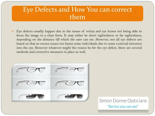
What is refractive error? • Refractive error is a focusing disorder of the eye • Most common cause of vision impairment • Over the age of 40 years, 22 per cent of the population has refractive error • It is correctable by wearing glasses or contact lenses or refractive laser surgery (selected cases)
Prevalence and risk factors of refractive error • All age groups can be affected by refractive error • People over the age of 40 should have regular eye tests to eliminate refractive error as a cause of any vision impairment • Family history of refractive error is a risk factor
Functional implications of refractive error Functional implications depend on the type of severity of refractive error: • long-sightedness (hyperopia) • difficulty seeing near objects • short-sightedness (myopia) • difficulty seeing things in the distance • astigmatism • blurred vision • presbyopia (age focus difficulty) • difficulty seeing near objects occurs from 40 and onwards
Types of Eye Hazards Flying objects Particles and dust Chemicals Harmful light radiation – ultraviolet, lasers, infrared 3
Sources of Eye Hazards Flying objects or particles in eye Grinding Sanding Sandblasting Blowdown Nail gun use Woodworking 4
Sources of Eye Hazards Chemical Hazards The most dangerous chemicals to the eyes are corrosive liquids. Examples include acids, lye, bleach, ammonia, sodium hydroxide and formaldehyde. Other chemicals can also be extremely irritating to the eyes. Some pesticides can be absorbed through the eyes and make you sick. 5
Sources of Eye Hazards Harmful light radiation Infrared from molten metal Ultraviolet from welding Laser 6
Types of Eye Protection Three Main Types Safety glasses goggles Face shields 8
Safety Glasses Side shields or wraparound required Must meet ANSI Z87.1 Standards for impact resistance Must be comfortable if worn for long periods 9