Download
1 / 1
10 likes | 74 Views
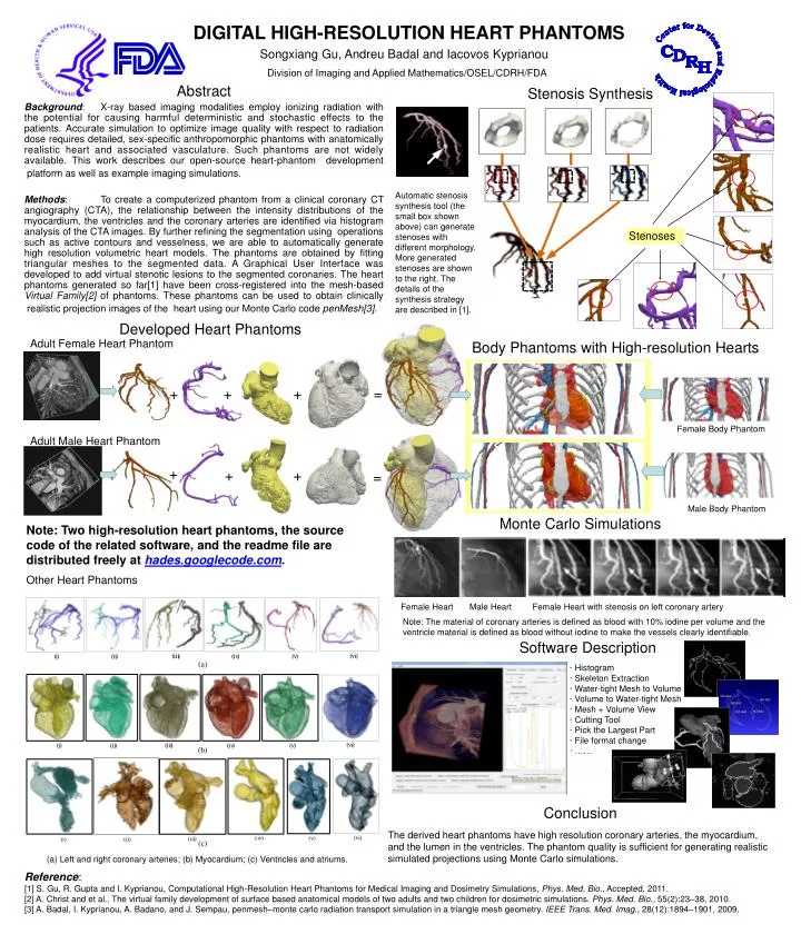
Songxiang Gu, Andreu Badal and Iacovos Kyprianou. Division of Imaging and Applied Mathematics/OSEL/CDRH/FDA. Stenosis Synthesis. DIGITAL HIGH-RESOLUTION HEART PHANTOMS.

E N D
Songxiang Gu, Andreu Badal and Iacovos Kyprianou Division of Imaging and Applied Mathematics/OSEL/CDRH/FDA Stenosis Synthesis DIGITAL HIGH-RESOLUTION HEART PHANTOMS Automatic stenosis synthesis tool (the small box shown above) can generate stenoses with different morphology. More generated stenoses are shown to the right. The details of the synthesis strategy are described in [1]. Stenoses Developed Heart Phantoms Adult Female Heart Phantom Body Phantoms with High-resolution Hearts + + + = Female Body Phantom Adult Male Heart Phantom + + + = Abstract Background: X-ray based imaging modalities employ ionizing radiation with the potential for causing harmful deterministic and stochastic effects to the patients. Accurate simulation to optimize image quality with respect to radiation dose requires detailed, sex-specific anthropomorphic phantoms with anatomically realistic heart and associated vasculature. Such phantoms are not widely available. This work describes our open-source heart-phantom development platform as well as example imaging simulations. Methods: To create a computerized phantom from a clinical coronary CT angiography (CTA), the relationship between the intensity distributions of the myocardium, the ventricles and the coronary arteries are identified via histogram analysis of the CTA images. By further refining the segmentation using operations such as active contours and vesselness, we are able to automatically generate high resolution volumetric heart models. The phantoms are obtained by fitting triangular meshes to the segmented data. A Graphical User Interface was developed to add virtual stenotic lesions to the segmented coronaries. The heart phantoms generated so far[1] have been cross-registered into the mesh-based Virtual Family[2] of phantoms. These phantoms can be used to obtain clinically realistic projection images of the heart using our Monte Carlo code penMesh[3]. Male Body Phantom Monte Carlo Simulations Note: Two high-resolution heart phantoms, the source code of the related software, and the readme file are distributed freely at hades.googlecode.com. Other Heart Phantoms Female Heart Male Heart Female Heart with stenosis on left coronary artery Note: The material of coronary arteries is defined as blood with 10% iodine per volume and the ventricle material is defined as blood without iodine to make the vessels clearly identifiable. Software Description · Histogram · Skeleton Extraction · Water-tight Mesh to Volume · Volume to Water-tight Mesh · Mesh + Volume View · Cutting Tool · Pick the Largest Part · File format change · …… Conclusion The derived heart phantoms have high resolution coronary arteries, the myocardium, and the lumen in the ventricles. The phantom quality is sufficient for generating realistic simulated projections using Monte Carlo simulations. (a) Left and right coronary arteries; (b) Myocardium; (c) Ventricles and atriums. Reference: [1] S. Gu, R. Gupta and I. Kyprianou, Computational High-Resolution Heart Phantoms for Medical Imaging and Dosimetry Simulations, Phys. Med. Bio., Accepted, 2011. [2] A. Christ and et al., The virtual family development of surface based anatomical models of two adults and two children for dosimetric simulations. Phys. Med. Bio., 55(2):23–38, 2010. [3] A. Badal, I. Kyprianou, A. Badano, and J. Sempau, penmesh–monte carlo radiation transport simulation in a triangle mesh geometry. IEEE Trans. Med. Imag., 28(12):1894–1901, 2009.