Download
1 / 34
350 likes | 664 Views
Perception of Motion, Depth and Form. David M. Waitzman, M.D., Ph.D. Discussion Question. Motion in Depth lecture points:

E N D
Perception of Motion, Depth and Form David M. Waitzman, M.D., Ph.D.
Discussion Question • Motion in Depth lecture points: • What are the two visual streams and what kinds of tasks are common to both. What tasks are different? If an object is moving toward you what portion of the hemisphere would be activated? What portion of the cortex would recognize the object? • Dorsal and Ventral • Common: Disparity Cells in visual (V1, V2, V4) AND extrastriate (MT) cortex respond to targets closer (orange) or further (blue) than fixation point (green) • Different: Motion versus Object recognition • Near: MST (looming objects) • Recognition: Area IT
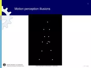
Phi or Beta Phenomenon The POINT of this Demonstration: The brain is wired to perceive motion even though in this demonstration none of the objects were actually moving
Perceived Motion: Analysis of Motion Occurs in a Separate Pathway Movement of an image on the retina or an eye movement (above) True movement or apparent (flashed) movement (right)
Receptive Fields in the Retina • Two types of ganglion cells: • on and off dependent upon the bipolar neurons • Center Surround structure of the receptive field described by Kuffler • Best activated by central illumination • Best inhibited by annular illumination
Different View of Center-Surround Organization: Parallel Pathways • Transformation of visual information is evident in the ganglion cells of the retina • X cells – sustained linear responses • Y cells – transient, excitatory non-linear responses
P and M Projections to LGN: Different Physiologic Channels • P cells in the retina (also known as midget ganglion cells) project to the parvocellular layers (3-6) of LGN • M cells in the retina (also known as parasol cells) project to the magnocellular (ventral most) layers (1-2) of the LGN • Intercalated layers are termed koniocellular (dustlike or tiny cells)
Comparison of P and M Pathways • P Parvocellular • Small Receptive Fields • Slow conduction to LGN • Sustained responses (X Like) • Sensitive to color • High contrast • M Magnocellular • Larger receptive fields • Rapid conduction to LGN • Transient responses (Y like) • Broadband color sensitivity • Low Contrast
Anatomic Segregation of Motion and Object Recognition Pathways • parvocellular: 4C“high acuity”; to blobs; to thin stripes, to V4 (color) • magnocellular: 4Ca “motion pathway”; to layer 4B of V1 and to V2 (area 18) and hence to Area MT
MT Solves the Aperture Problem Top : Grating moving in any of 3 apertures has the same apparent motion Bottom: In MT two edge detecting neurons are linked to permit identification of the overall motion of the entire object
Plaid Motion: V1 versus Area MT Neurons in V1 encode the direction of individual components of motion (top) MT neurons encode the perceived speed and direction of the moving visual stimulus (below)
Confirmation of Different Pathways from Clinical Syndromes Type of Agnosia Symptoms Can’t name use or recognize real objects Doesn’t recognize drawn objects Can’t recognize faces Doesn’t associate a color with an object Can’t name colors Can’t distinguish hues Loss of stereoscopic vision Can’t see objects moving • Object agnosia • Agnosia for drawings • Prosopagnosia • Color Agnosia • Color anomia • Achromatopsia • Visual spatial agnosia • Movement agnosia
Dorsal and Ventral Streams • Dorsal Stream: Where: Motion • MT projects to MST • MST projects to inferior parietal • lobule 7a and to the FEF • Ventral Stream: What : Form and Color
Smooth Pursuit: Characteristics Purpose: Track visual stimuli moving at < 100/s Stimulus: Step-Ramp
Smooth Pursuit: Characteristics Despite a target appearance on the contralateral side, initial pursuit velocity is in the direction of smooth motion
Area MT Physiology • MT neurons encode visual stimulus motion (discharge is reduced when target is blanked)
MT Lesion: Scotoma for Motion • Scotoma (i.e., blindness) for motion in a specific portion of the contralateral visual hemifield. • Saccades to moving targets in the blind area are inaccurate, e.g., note the overshoot of the leftward saccade at down arrow • Monkeys cannot estimate the speed of the target until it is outside the “motion blind” area • Saccades to stationary targets are normometric
Area MST: Perception of Eye Motion • Neurons in area MST receive a signal of current eye and head velocity (A) • large visual fields • can estimate heading and target motion by eliminating self motion • Discharge not reduced when visual stimulus is blanked or stabilized (B)
MST Lesions: Loss of Eye Speed Toward the Ipsilateral Side Two deficits: • retinotopic deficit: cannot estimate speed in a region of contralateral visual hemifield • unidirectional loss of smooth pursuit toward the side of the lesion REGARDLESS of which visual hemifield the stimulus is presented
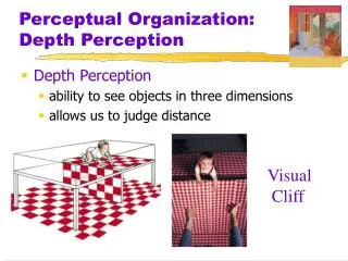
Monocular Cues for Depth Perception • Familiar Size • Occlusion • Linear perspective • Size perspective • Distribution of shadows and illumination • Motion Parallax • And…..
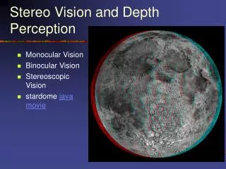
Binocular Cues for Depth Perception: Disparity Tuning A binocular disparity signal arises in V1 (Hubel and Wiesel) Cells in visual (V1, V2, V4) AND extrastriate (MT) cortex respond to targets closer (orange) or further (blue) than fixation point (green) Both dorsal and ventral pathways have disparity tuning
Dorsal Pathway (Area MT): Direction and Disparity Tuning Tuning for targets closer or further from the monkey (near and far response patches) Newsome et al, J. Neurosci. 1999
Ventral Pathway (V4): Binocular Disparity • Tuning in depth • Dorsal and Ventral Streams NOT Segregated for Disparity (a common feature)
Color and Form Are Major Differences in Ventral Path • Human subjects undergo PET scan to demonstrate motion regions (left column) and color regions (right column) corresponding with monkey area MT and V4 respectively
Illusory Contours in V2 Neurons • Receptive field of the neuron is the ellipse • Strong activation when an edge passes over the receptive field • Persistent activation when an illusory contour passes over receptive field (2) • No response when the edge is partial (3 and 4)
V4 Neurons Sense Gaze position (not movement) and Relative Size • View objects at 3 different distances and at 9 different gaze positions
Spike rasters of the stimulus (S) period for each of the 27 experimental conditions • The center figure represents the mean firing rates for this cell during both the fixation period (FO, blue bars) and the stimulation period (S, red bars) • Leftmost graphs show the mean firing rate data during the stimulation period using an interpolated color plot. Each of the planes represents a monitor viewing distance.
V4 Neurons: Complex forms, size, orientation, color, but not Faces • On the right, contour plot of the S (size) regions (WHITE) is superimposed on a color-coded map of orientation preference. Color saturation within this map indicates orientation selectivity. Unlike in V2, S regions in V4 are not limited to unoriented regions, but can span iso-orientation domains.
Inferotemporal cortex respond to form, color, and faces Cells increase in complexity of object recognition, but lose the ability to spatially localize objects (no gaze direction sensitivity) Face recognition is present
Temporal Lesions: Loss of Object Recognition and Prosopagnosia Regions for face recognition have been demonstrated by fMRI studies include the FFA and PPA (temporal lobe) and overlap with object recognition regions Haxby et al, Science 293: 2425-2430 , 2001. Neural basis of prosopagnosia… N. Hadjikhani and B. de Gelder, Human Brain Mapping, 16(3):176-182, 2002.
Visual Attention: Solves the binding problem • The activity of V4 neurons is greatly enhanced by selective attention to the color of the matching stimulus (red) • Significance for the monkey is greatly increased for the matched (above) then the unmatched trials (below)
Attention causes activation of a parieto-frontal pathway • Lack of attention to visual stimuli produced NO activation of the cortex (A)! • With attention to the peripheral target with discrimination, activation of a parieto-frontal pathway either directly (B) or in anticipation of target appearance (C).