Download
1 / 1
10 likes | 137 Views
APP as a Dependence Receptor. ABSTRACT. Screening for the ApoE-APP-Tau complex inhibitors: Relevance for Alzheimer’s Disease. Gabriellee Cailing , Sonia Flores, Alex Patent , Varghese John, Rammohan V. Rao* and Dale E. Bredesen* Buck Institute for Research on Aging, Novato, CA.

E N D
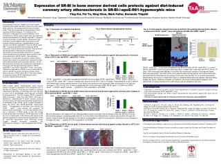
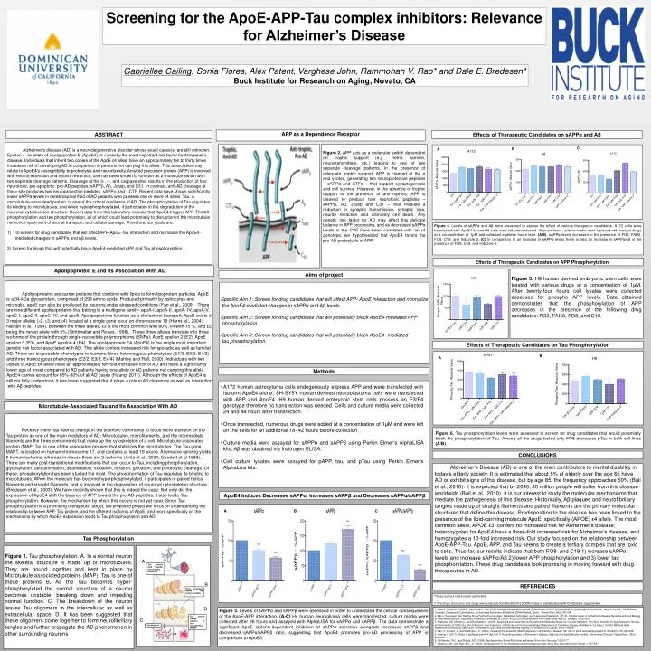
APP as a Dependence Receptor ABSTRACT Screening for the ApoE-APP-Tau complex inhibitors: Relevance for Alzheimer’s Disease Gabriellee Cailing, Sonia Flores, Alex Patent, Varghese John, Rammohan V. Rao* and Dale E. Bredesen* Buck Institute for Research on Aging, Novato, CA Effects of Therapeutic Candidates on sAPPα and Aβ C B A Figure 2. APP acts as a molecular switch dependent on trophic support (e.g., netrin, laminin, neurotransmitters, etc.) leading to one of two separate cleavage patterns. In the presence of adequate trophic support, APP is cleaved at the α and γ sites, generating two neuroprotective peptides – sAPPαand CTFα – that support synaptogenesis and cell survival. However, in the absence of trophic support or the presence of anti-trophins, APP is cleaved to produce four neurotoxic peptides – sAPPβ, Aβ, Jcasp and C31 – that mediate a reduction in synaptic transmission, synaptic loss, neurite retraction and ultimately cell death. Any genetic risk factor for AD may affect this delicate balance in APP processing, and as decreased sAPPα levels in the CSF have been correlated with an ε4 genotype, we hypothesized that ApoE4 favors the pro-AD proteolysis of APP. • Alzheimer’s disease (AD) is a neurodegenerative disorder whose exact cause(s) are still unknown. Εpsilon 4, an allele of apolipoprotein E (ApoE4), is currently the most important risk factor for Alzheimer’s disease. Individuals that inherit two copies of the ApoE ε4 allele have an approximately ten to thirty times increased risk of developing AD in comparison to persons not carrying this allele. This association may relate to ApoE4’s susceptibility to proteolysis and neurotoxicity. Amyloid precursor protein (APP) is involved with neurite extension and neurite retraction, and has been shown to function as a molecular switch with two separate cleavage patterns. Cleavage at the -, -, and caspase sites results in the production of four neurotoxic, pro-apoptotic, pro-AD peptides: sAPP, A, Jcasp, and C31. In contrast, anti-AD cleavage at the -site produces two neuroprotective peptides: sAPP and CTF. Recent data have shown significantly lower sAPPα levels in cerebrospinal fluid of AD patients who possess one or more ε4 allele. Tau, a microtubule-associated protein, is one of the critical mediators of AD. The phosphorylation of Tau regulates its binding to microtubules, and when hyperphosphorylated, it participates in the degradation of the neuronal cytoskeleton structure. Recent data from this laboratory indicate that ApoE4 triggers APP-Thr668 phosphorylation and tau phosphorylation, all of which could lead potentially to disruption of the microtubule network, impairment of axonal transport, and cellular damage. Therefore, our goals are: • To screen for drug candidates that will affect APP-ApoE-Tau interaction and normalize the ApoE4- mediated changes in sAPPα and Aβ levels. • 2) Screen for drugs that will potentially block ApoE4-mediated APP and Tau phosphorylation. Figure 4. Levels of sAPPα and Aβ were measured to assess the effect of various therapeutic candidates. A172 cells were transfected with ApoE4 or and H9 cells were left untransfected. After six hours, culture media were replaced with various drugs at a concentration of 1μM and collected eighteen hours later. (A/B). sAPPα levels increased in the presence of FO3, FAH3, FO8, C19, and Indazole 2. (C) In comparison to an increase in sAPPα levels there is also an increase in sAPPα/Aβ in the presence of FO8, C19, and Indazole 2. Effects of Therapeutic Candidates on APP Phosphorylation Figure 5. H9 human derived embryonic stem cells were treated with various drugs at a concentration of 1μM. After twenty-four hours cell lysates were collected assessed for phosphoAPP levels. Data obtained demonstrates that the phosphorylation of APP decreases in the presence of the following drug candidates: FO3, FAH3, FO8, and C19. Aims of project Specific Aim 1: Screen for drug candidates that will affect APP- ApoE interaction and normalize the ApoE4-mediated changes in sAPPα and Aβ levels. Specific Aim 2: Screen for drug candidates that will potentially block ApoE4-mediated APP phosphorylation. Specific Aim 3: Screen for drug candidates that will potentially block ApoE4- mediated tau phosphorylation. Apolipoprotein E and Its Association With AD Apolipoproteins are carrier proteins that combine with lipids to form lipoprotein particles.ApoE is a 34-kDa glycoprotein, comprised of 299 amino acids. Produced primarily by astrocytes and microglia, apoE can also be produced by neurons under stressed conditions (Fan et al., 2009). There are nine different apolipoproteins that belong to a multigene family- apoA-I, apoA-II, apoA- IV, apoA-V, apoC-I, apoC-II, apoC- IV, and apoE. Apolipoproteins function as a cholesterol transport. ApoE exists in 3 major alleles (2, 3, and 4) located at a single gene locus on chromosome 19 (Harris et., 2004; Nathan et al., 1994). Between the three alleles, ε3 is the most common with 80%, ε4 with 15 %, and ε2 being the rarest allele with 5% (Strittmatter and Roses, 1996). These three alleles translate into three isoforms of the protein through single-nucleotide polymorphisms (SNPs): ApoE epsilon 2 (E2), ApoE epsilon 3 (E3), and ApoE epsilon 4 (E4). The apolipoprotein E4 (ApoE4) is the single most important genetic risk factor associated with AD. This allele confers increased risk for sporadic as well as familial AD. There are six possible phenotypes in humans: three heterozygous phenotypes (E4/3, E3/2, E4/2) and three homozygous phenotypes (E2/2, E3/3, E4/4) (Mahley and Rall, 2000). Individuals with two copies of ApoE ε4 allele have an approximately ten-fold increased rick of AD and have a significantly lower age of onset compared to AD patients having one allele or AD patients not carrying this allele. ApoE4 carries account for 65%-80% of all AD cases (Huang, 2011). Although the effects of ApoE4 is still not fully understood, it has been suggested that it plays a role in Aβ clearance as well as interaction with Aβ peptides. Effects of Therapeutic Candidates on Tau Phosphorylation B A Methods • A172 human astrocytoma cells endogenously express APP and were transfected with isoform ApoE4 alone. SH-SY5Y human derived neuroblastoma cells were transfected with APP and ApoE4. H9 human derived embryonic stem cells possess an E3/E4 genotype therefore no transfection was needed. Cells and culture media were collected 24and 48 hours after transfection. • Once transfected, numerous drugs were added at a concentration of 1μM and were left on the cells for an additional 18- 42 hours before collection. • Culture media were assayed for sAPPα and sAPPβ using Perkin Elmer’s AlphaLISA kits. Aβ was obtained via Invitrogen ELISA. • Cell culture lysates were assayed for pAPP, tau, and pTau using Perkin Elmer’s AlphaLisa kits. Microtubule-Associated Tau and Its Association With AD Recently there has been a change in the scientific community to focus more attention on the Tau protein as one of the main mediators of AD. Microtubules, microfilaments, and the intermediate filaments are the three components that make up the cytoskeleton of a cell. Microtubule-associated protein (MAP) Tau is one of the associated proteins that stabilizes the microtubules. The Tau gene, MAPT, is located on human chromosome 17, and contains at least 16 exons. Alternative splicing yields 6 human isoforms, whereas in mouse there are 2 isoforms (Avila et al., 2004; Goedert et al 1989). There are many post-translational modifications that can occur to Tau, including phosphorylation, glycosylation, ubiquitinylation, deamidation, oxidation, nitration, glycation, and proteolytic cleavage. Of these, phosphorylation has been studied the most. The phosphorylation of Tau regulates its binding to microtubules. When the molecule has become hyperphosphorylated, it participates in paired helical filaments and straight filaments, and is involved in the degradation of neuronal cytoskeleton structure (Bredesen et al., 2003). We have recently shown that this is indeed the case. Not only did the expression of ApoE4 shift the balance of APP toward the pro-AD peptides, it also led to Tau phosphorylation. However, the mechanism by which this occurs is not yet clear. Since Tau phosphorylation is a promising therapeutic target, the proposed project will focus on understanding the relationship between APP, Tau protein, and the different isoforms of ApoE, and more specifically on the mechanisms by which ApoE4 expression leads to Tau phosphorylation and AD. Figure 6. Tau phosphorylation levels were assessed to screen for drug candidates that would potentially block the phosphorylation of Tau. Among all the drugs tested only FO8 decreases pTau in both cell lines (A/B) CONCLUSIONS Alzheimer’s Disease (AD) is one of the main contributors to mental disability in today’s elderly society. It is estimated that about 5% of elderly over the age 65 have AD or exhibit signs of this disease, but by age 85, the frequency approaches 50% (Bali et al., 2010). It is expected that by 2040, 80 million people will suffer from this disease worldwide (Bali et al., 2010). It is our interest to study the molecular mechanisms that mediate the pathogenesis of this disease. Historically, Aβ plaques and neurofibrillary tangles made up of straight filaments and paired filaments are the primary molecular structures that define this disease. Predisposition to the disease has been linked to the presence of the lipid-carrying molecule ApoE, specifically (APOE) ε4 allele.The most common allele, APOE ε3, confers no increased risk for Alzheimer’s disease; heterozygotes for ApoE4 have a three-fold increased risk for Alzheimer’s disease, and homozygotes a 10-fold increased risk.Our study focused on the relationship between ApoE-APP-Tau. ApoE, APP, and Tau seems to create a tertiary complex that are toxic to cells. Thus far, our results indicate that both FO8 and C19 1) increase sAPPα levels and increase sAPPα/Aβ 2) lower APP phosphorylation and 3) lower tau phosphorylation. These drug candidates look promising in moving forward with drug therapeutics in AD. ApoE4 Induces Decreases sAPPα, Increases sAPPβ and Decreases sAPPα/sAPPβ Tau proteins hold together microtubules inside cell REFERENCES A C B 1. Avila J, Lucas JJ, Perez M, Hernandez F. Centro de Biología Molecular2004 Role of tau protein in both physiological and pathological conditions. "Severo Ochoa", Facultad de Ciencias,.Campus de Cantoblanco, Universidad Autónoma de Madrid, 28049 Madrid, Spain. Physiol Rev.;84(2):361-84 2. Bali J, Saoussen Ben Halima, Boas Felmy, Zoe Goodger, Sebastian Zurbriggen, and Lawrence Rajendran. (2010). Cellular basis of Alzheimer’s disease.Systems and Cell Biology of Neurodegeneration, Psychiatry Research, University of Zürich, 8008 Zurich, Switzerland Ann Indian Acad Neurol. (Suppl2): S89–S93 3. Bredesen DE, Mehlen p., and Rabizadeh S. (2003). Apoptosis and Dependence Receptors: A Molecular Basis for Cellular Addiction. The Buck Institute for Age Research, Novato, and University of California, San Francisco, San Francisco, California; and Centre Genetique Moleculaire et Cellulaire, Equipe Labellise´e “La Ligue,” Centre National de la Recherche Scientifique UMR5534, University of Lyon, and the International Agency For Research in Cancer, Lyon, France 4. Fan, J., Donkin, J., and Wellington, C. (2009). Greasing the wheels of Abeta clearance in Alzheimer's disease: the role of lipids and apolipoprotein E. Biofactors 35, 239-248. 5. Huang, Y. (2011). Roles of apolipoprotein E4 (ApoE4) in the pathogenesis of Alzheimer's disease: lessons from ApoE mouse models. Biochemical Society Transactions, 39(4), 924-932. 6. Strittmatter, W.J., and Roses, A.D. (1996). Apolipoprotein E and Alzheimer's disease. Annu Rev Neurosci 19, 53-77. 7. Mahley, R.W., and Rall, S.C., Jr. (2000). Apolipoprotein E: far more than a lipid transport protein. Annu Rev Genomics Hum Genet 1, 507-537. Tau Phosphorylation Figure 1. Tau phosphorylation. A. In a normal neuron the skeletal structure is made up of microtubules. They are bound together and kept in place by Microtubule associated proteins (MAP); Tau is one of these proteins B. As the Tau becomes hyper-phosphorylated the normal structure of a neuron becomes unstable, breaking down and impeding normal function. C. The breakdown of the neuron leaves Tau oligomers in the intercellular as well as extracellular space. D. It has been suggested that these oligomers come together to form neurofibrillary tangles and further propagate the AD phenomenon in other surrounding neurons A B Extra phosphate makes Tau protein unstable *These authors share senior authorship ♯The drugs chosen for this study were selected from the BUCK’s ADDN library in collaboration with Dr. Barbara Jagodzinska. D Figure 3. Levels of sAPPα and sAPPβ were assessed in order to understand the cellular consequences of the ApoE-APP interaction. (A-C) H4 human neuroglioma cells were transfected, culture media were collected after 36 hours and assayed with AlphaLISA for sAPPα and sAPPβ. The data demonstrate a significant ApoE isoform-dependent inhibition of sAPPα secretion alongside increased sAPPβ and decreased sAPPα/sAPPβ ratio, suggesting that ApoE4 promotes pro-AD processing of APP in comparison to ApoE3. Tau protein clumps together to form neurofibrillary tangles C Tau oligomer