Download
1 / 56
560 likes | 708 Views
Osmoregulation. (this one’s a real pisser). Overview: A Balancing Act. Physiological systems of animals operate in a fluid environment Relative concentrations of water and solutes must be maintained within fairly narrow limits

E N D
Osmoregulation (this one’s a real pisser)
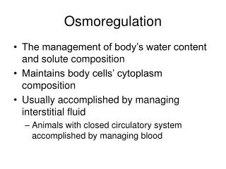
Overview: A Balancing Act • Physiological systems of animals operate in a fluid environment • Relative concentrations of water and solutes must be maintained within fairly narrow limits • Osmoregulationregulates solute concentrations and balances the gain and loss of water • Freshwater animals show adaptations that reduce water uptake and conserve solutes • Desert and marine animals face desiccating environments that can quickly deplete body water • Excretion gets rid of nitrogenous metabolites and other waste products
Osmosis and Osmolarity • Cells require a balance between osmotic gain and loss of water • Osmolarity, the solute concentration of a solution, determines the movement of water across a selectively permeable membrane • If two solutions are isoosmotic, the movement of water is equal in both directions • If two solutions differ in osmolarity, the net flow of water is from the hypoosmotic to the hyperosmotic solution
Fig. 44-2 Selectively permeable membrane Solutes Net water flow Water Hypoosmotic side Hyperosmotic side
Osmotic Challenges • Osmoconformers, consisting only of some marine animals, are isoosmotic with their surroundings and do not regulate their osmolarity • Osmoregulators expend energy to control water uptake and loss in a hyperosmotic or hypoosmotic environment
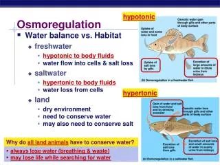
Marine Animals • Most marine invertebrates are osmoconformers • Most marine vertebrates and some invertebrates are osmoregulators • Marine bony fishes are hypoosmotic to sea water • They lose water by osmosis and gain salt by diffusion and from food • They balance water loss by drinking seawater and excreting salts
Fig. 44-4 Gain of water and salt ions from food Osmotic water loss through gills and other parts of body surface Uptake of water and some ions in food Excretion of salt ions from gills Uptake of salt ions by gills Osmotic water gain through gills and other parts of body surface Gain of water and salt ions from drinking seawater Excretion of salt ions and small amounts of water in scanty urine from kidneys Excretion of large amounts of water in dilute urine from kidneys (a) Osmoregulation in a saltwater fish (b) Osmoregulation in a freshwater fish
Fig. 44-5 100 µm 100 µm (b) Dehydrated tardigrade (a) Hydrated tardigrade
Fig. 44-6 Water balance in a kangaroo rat (2 mL/day) Water balance in a human (2,500 mL/day) Ingested in food (0.2) Ingested in food (750) Ingested in liquid (1,500) Water gain (mL) Derived from metabolism (250) Derived from metabolism (1.8) Feces (0.09) Feces (100) Water loss (mL) Urine (1,500) Urine (0.45) Evaporation (1.46) Evaporation (900)
Transport Epithelia in Osmoregulation • Animals regulate the composition of body fluid that bathes their cells • Transport epithelia are specialized epithelial cells that regulate solute movement • They are essential components of osmotic regulation and metabolic waste disposal • They are arranged in complex tubular networks • An example is in salt glands of marine birds, which remove excess sodium chloride from the blood
Fig. 44-7 EXPERIMENT Nasal salt gland Ducts Nostril with salt secretions
Fig. 44-8 Vein Artery Secretory tubule Secretory cell Salt gland Capillary Secretory tubule Transport epithelium NaCl NaCl Direction of salt movement Central duct Blood flow Salt secretion (b) (a)
Fig. 44-9 Proteins Nucleic acids Amino acids Nitrogenous bases Amino groups Most aquatic animals, including most bony fishes Mammals, most amphibians, sharks, some bony fishes Many reptiles (including birds), insects, land snails Ammonia Uric acid Urea
Ammonia • Animals that excrete nitrogenous wastes as ammonia need lots of water • They release ammonia across the whole body surface or through gills
Urea • The liver of mammals and most adult amphibians converts ammonia to less toxic urea • The circulatory system carries urea to the kidneys, where it is excreted • Conversion of ammonia to urea is energetically expensive; excretion of urea requires less water than ammonia
Uric Acid • Insects, land snails, and many reptiles, including birds, mainly excrete uric acid • Uric acid is largely insoluble in water and can be secreted as a paste with little water loss • Uric acid is more energetically expensive to produce than urea
Excretory Processes • Most excretory systems produce urine by refining a filtrate derived from body fluids • Key functions of most excretory systems: • Filtration: pressure-filtering of body fluids • Reabsorption: reclaiming valuable solutes • Secretion: adding toxins and other solutes from the body fluids to the filtrate • Excretion: removing the filtrate from the system
Survey of Excretory Systems • Systems that perform basic excretory functions vary widely among animal groups • They usually involve a complex network of tubules
Protonephridia • A protonephridium is a network of dead-end tubules connected to external openings • The smallest branches of the network are capped by a cellular unit called a flame bulb • These tubules excrete a dilute fluid and function in osmoregulation
Fig. 44-11 Nucleus of cap cell Cilia Flame bulb Interstitial fluid flow Opening in body wall Tubule Tubules of protonephridia Tubule cell
Metanephridia • Each segment of an earthworm has a pair of open-ended metanephridia • Metanephridia consist of tubules that collect coelomic fluid and produce dilute urine for excretion
Fig. 44-12 Coelom Capillary network Components of a metanephridium: Internal opening Collecting tubule Bladder External opening
Malpighian Tubules • In insects and other terrestrial arthropods, Malpighian tubules remove nitrogenous wastes from hemolymph and function in osmoregulation • Insects produce a relatively dry waste matter, an important adaptation to terrestrial life
Fig. 44-13 Digestive tract Rectum Hindgut Intestine Midgut (stomach) Malpighian tubules Salt, water, and nitrogenous wastes Feces and urine Rectum Reabsorption HEMOLYMPH
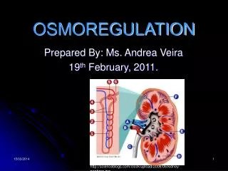
Kidneys • Kidneys, the excretory organs of vertebrates, function in both excretion and osmoregulation • The mammalian excretory system centers on paired kidneys, which are also the principal site of water balance and salt regulation • Each kidney is supplied with blood by a renal artery and drained by a renal vein • Urine exits each kidney through a duct called the ureter • Both ureters drain into a common urinary bladder, and urine is expelled through a urethra
Fig. 44-14ab Renal medulla Posterior vena cava Renal cortex Renal artery and vein Kidney Renal pelvis Aorta Ureter Urinary bladder Ureter Urethra Section of kidney from a rat (a) Excretory organs and major associated blood vessels (b) Kidney structure 4 mm
Fig. 44-14a Posterior vena cava Renal artery and vein Kidney Aorta Ureter Urinary bladder Urethra (a) Excretory organs and major associated blood vessels
The mammalian kidney has two distinct regions: an outer renal cortex and an inner renal medulla
Fig. 44-14b Renal medulla Renal cortex Renal pelvis Ureter Section of kidney from a rat (b) Kidney structure 4 mm
The nephron, the functional unit of the vertebrate kidney, consists of a single long tubule and a ball of capillaries called the glomerulus • Bowman’s capsule surrounds and receives filtrate from the glomerulus
Fig. 44-14c Juxtamedullary nephron Cortical nephron Renal cortex Collecting duct Renal medulla To renal pelvis (c) Nephron types
Fig. 44-14d Glomerulus Afferent arteriole from renal artery Bowman’s capsule 10 µm SEM Proximal tubule Peritubular capillaries Efferent arteriole from glomerulus Distal tubule Branch of renal vein Collecting duct Descending limb Loop of Henle Ascending limb Vasa recta (d) Filtrate and blood flow
Filtration of the Blood • Filtration occurs as blood pressure forces fluid from the blood in the glomerulus into the lumen of Bowman’s capsule • Filtration of small molecules is nonselective • The filtrate contains salts, glucose, amino acids, vitamins, nitrogenous wastes, and other small molecules
Pathway of the Filtrate • From Bowman’s capsule, the filtrate passes through three regions of the nephron: the proximal tubule, the loop of Henle, and the distal tubule • Fluid from several nephrons flows into a collecting duct, all of which lead to the renal pelvis,which is drained by the ureter • Cortical nephrons are confined to the renal cortex, while juxtamedullary nephrons have loops of Henle that descend into the renal medulla
Blood Vessels Associated with the Nephrons • Each nephron is supplied with blood by an afferent arteriole, a branch of the renal artery that divides into the capillaries • The capillaries converge as they leave the glomerulus, forming an efferent arteriole • The vessels divide again, forming the peritubular capillaries, which surround the proximal and distal tubules
Concept 44.4: The nephron is organized for stepwise processing of blood filtrate • The mammalian kidney conserves water by producing urine that is much more concentrated than body fluids
Descending Limb of the Loop of Henle • Reabsorption of water continues through channels formed by aquaporin proteins • Movement is driven by the high osmolarity of the interstitial fluid, which is hyperosmotic to the filtrate • The filtrate becomes increasingly concentrated
From Blood Filtrate to Urine: A Closer Look Proximal Tubule • Reabsorption of ions, water, and nutrients takes place in the proximal tubule • Molecules are transported actively and passively from the filtrate into the interstitial fluid and then capillaries • Some toxic materials are secreted into the filtrate • The filtrate volume decreases
Ascending Limb of the Loop of Henle • In the ascending limb of the loop of Henle, salt but not water is able to diffuse from the tubule into the interstitial fluid • The filtrate becomes increasingly dilute
Distal Tubule • The distal tubule regulates the K+ and NaCl concentrations of body fluids • The controlled movement of ions contributes to pH regulation
Collecting Duct • The collecting duct carries filtrate through the medulla to the renal pelvis • Water is lost as well as some salt and urea, and the filtrate becomes more concentrated • Urine is hyperosmotic to body fluids
Fig. 44-15 Proximal tubule Distal tubule NaCl Nutrients H2O HCO3– H2O K+ HCO3– NaCl H+ H+ NH3 K+ Filtrate CORTEX Loop of Henle NaCl H2O OUTER MEDULLA NaCl NaCl Collecting duct Key Urea NaCl Active transport H2O INNER MEDULLA Passive transport
The Two-Solute Model • In the proximal tubule, filtrate volume decreases, but its osmolarity remains the same • The countercurrent multiplier system involving the loop of Henle maintains a high salt concentration in the kidney • This system allows the vasa recta to supply the kidney with nutrients, without interfering with the osmolarity gradient • Considerable energy is expended to maintain the osmotic gradient between the medulla and cortex
Fig. 44-16-1 Osmolarity of interstitial fluid (mOsm/L) 300 300 300 300 H2O CORTEX 400 400 H2O H2O H2O OUTER MEDULLA 600 600 H2O H2O 900 900 Key H2O Active transport INNER MEDULLA 1,200 Passive transport 1,200
Fig. 44-16-2 Osmolarity of interstitial fluid (mOsm/L) 300 300 100 300 100 300 H2O NaCl CORTEX 200 400 400 NaCl H2O H2O NaCl H2O NaCl OUTER MEDULLA 600 400 600 NaCl H2O NaCl H2O 900 900 700 Key NaCl H2O Active transport INNER MEDULLA 1,200 Passive transport 1,200
Fig. 44-16-3 Osmolarity of interstitial fluid (mOsm/L) 300 300 100 300 100 300 300 H2O H2O NaCl CORTEX 400 200 400 400 H2O NaCl H2O NaCl H2O H2O NaCl NaCl H2O NaCl H2O OUTER MEDULLA 600 400 600 600 H2O NaCl H2O Urea NaCl H2O H2O 900 900 700 Urea Key NaCl H2O H2O Active transport INNER MEDULLA Urea 1,200 1,200 Passive transport 1,200
The Renin-Angiotensin-Aldosterone System • The renin-angiotensin-aldosterone system (RAAS) is part of a complex feedback circuit that functions in homeostasis • A drop in blood pressure near the glomerulus causes the juxtaglomerular apparatus (JGA) to release the enzyme renin • Renin triggers the formation of the peptide angiotensin II