Download
1 / 43
440 likes | 1.15k Views
Principal Points. Cloning is the making of many copies of a segment of DNACloning vectors range from plasmids to artificial chromosomes to virusesGenomic libraries are collections of clones that contain at least one copy of every DNA sequence in an organism's genomecDNA made from mRNA represent eukaryotic genes without intronsIdentification of clones can be done using cDNA probes, antibodies, complementation of mutations, and sequencingThe PCR is a procedure for amplifying a specific segment of DNA.

E N D
1. Chapter 8: Recombinant DNA Technology Linnea Fletcher Ph.D.
BIOL 2316
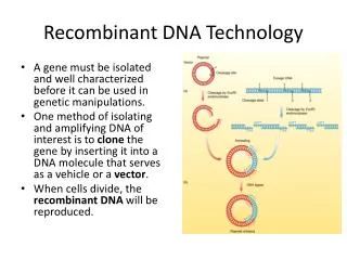
2. Principal Points Cloning is the making of many copies of a segment of DNA
Cloning vectors range from plasmids to artificial chromosomes to viruses
Genomic libraries are collections of clones that contain at least one copy of every DNA sequence in an organism�s genome
cDNA made from mRNA represent eukaryotic genes without introns
Identification of clones can be done using cDNA probes, antibodies, complementation of mutations, and sequencing
The PCR is a procedure for amplifying a specific segment of DNA
3. DNA fragments separated by gel electrophoresis and visualized under UV light. DNA fragments separated by gel electrophoresis and visualized under UV light.
4. What are the 3 general steps used to clone DNA?
Isolate DNA from an organism
Cut the organismal DNA and the vector with restriction enzymes making recombinant DNA
Introduce the recombinant DNA into a host
Restriction Enzymes
Recognize a specific site (generally a pallidromic sequence)
Produce overhangs or straight cuts
Naturally found in bacteria, they protect against viruses and foreign DNA
More than 400 enzymes have been isolated
Named for the organism they from which they are isolated
The first letter is that of the genus and the 2nd and 3rd are from the species
5. Figure 8.1 Restriction site in DNA, showing symmetry of the sequence around the center point. The sequence is a palindrome, reading the same from left to right (5�-to-3�) on the top strand (GAATTC, here) as it does from right to left (5�-to-3�) on the bottom strand. Shown is the restriction site for EcoRI. Figure 8.1 Restriction site in DNA, showing symmetry of the sequence around the center point. The sequence is a palindrome, reading the same from left to right (5�-to-3�) on the top strand (GAATTC, here) as it does from right to left (5�-to-3�) on the bottom strand. Shown is the restriction site for EcoRI.
7. Figure 8.2 Examples of how restriction enzymes cleave DNA. (a) SmaI results in blunt ends. (b) BamHI results in 5� overhanging (�sticky�) ends. (c) PstI results in 3� overhanging (�sticky�) ends. Figure 8.2 Examples of how restriction enzymes cleave DNA. (a) SmaI results in blunt ends. (b) BamHI results in 5� overhanging (�sticky�) ends. (c) PstI results in 3� overhanging (�sticky�) ends.
8. Figure 8.3 Cleavage of DNA by the restriction enzyme EcoRI. EcoRI makes staggered, symmetrical cuts in DNA, leaving �sticky� ends. A DNA fragment with a sticky end produced by EcoRI digestion can bind by complementary base pairing (anneal) to any other DNA fragment with a sticky end produced by EcoRI cleavage. The gaps can then be sealed by DNA ligase. Figure 8.3 Cleavage of DNA by the restriction enzyme EcoRI. EcoRI makes staggered, symmetrical cuts in DNA, leaving �sticky� ends. A DNA fragment with a sticky end produced by EcoRI digestion can bind by complementary base pairing (anneal) to any other DNA fragment with a sticky end produced by EcoRI cleavage. The gaps can then be sealed by DNA ligase.
9. What types of nucleic acids are used as cloning vectors?
What are some elements that are necessary for a plasmid cloning vector and in what ways is the pUC19 vector especially useful for cloning DNA?
Why must a cloning plasmid be recircularized before transforming a host bacterium?
Describe how insertional mutagenesis is used to distinguish nonrecombinant plasmid from recombinant plasmid when screening transformed hosts.
What are some elements that are necessary for a shuttle plasmid cloning vector?
What are some elements that are necessary for an expression cloning vector, a YAC, and a BAC?
Compare the size of DNA insert that can be carried by a plasmid, a YAC, and a BAC.
10. Figure 8.4 The plasmid cloning vector pUC19. This plasmid has an origin of replication (ori), an ampR selectable marker, and a polylinker located within part of the ?-galactosidase gene lacZ+. Figure 8.4 The plasmid cloning vector pUC19. This plasmid has an origin of replication (ori), an ampR selectable marker, and a polylinker located within part of the ?-galactosidase gene lacZ+.
11. Figure 8.5 Insertion of a piece of DNA into the plasmid cloning vector pUC19 to produce a recombinant DNA molecule. The vector pUC19 contains several unique restriction enzyme sites localized in a polylinker that are convenient for constructing recombinant DNA molecules. The insertion of a DNA fragment into the polylinker disrupts part of the ?-galactosidase (lacZ+) gene, leading to nonfunctional ? -galactosidase in E. coli. The blue�white color selection test described in the text can be used to select for vectors with or without inserts. Figure 8.5 Insertion of a piece of DNA into the plasmid cloning vector pUC19 to produce a recombinant DNA molecule. The vector pUC19 contains several unique restriction enzyme sites localized in a polylinker that are convenient for constructing recombinant DNA molecules. The insertion of a DNA fragment into the polylinker disrupts part of the ?-galactosidase (lacZ+) gene, leading to nonfunctional ? -galactosidase in E. coli. The blue�white color selection test described in the text can be used to select for vectors with or without inserts.
12. Figure 8.6 Example of a yeast artificial chromosome (YAC) cloning vector. A YAC vector contains a yeast telomere (TEL) at each end, a yeast centromere sequence (CEN), a yeast selectable marker for each arm (here, TRP1 and URA3), a sequence that allows autonomous replication in yeast (ARS), and restriction sites for cloning. Figure 8.6 Example of a yeast artificial chromosome (YAC) cloning vector. A YAC vector contains a yeast telomere (TEL) at each end, a yeast centromere sequence (CEN), a yeast selectable marker for each arm (here, TRP1 and URA3), a sequence that allows autonomous replication in yeast (ARS), and restriction sites for cloning.
13. What types of DNA inserts are found in genomic libraries, chromosomal libraries, and a cDNA library?
Describe the steps in constructing a genomic library.
What strategies can be used to ensure that the DNA inserts for a genomic library are especially large?
How can you avoid �gaps� in a genomic library?
How can a chromosome be segregated for constructing a chromosomal library?
14. What are some advantages of constructing a cDNA library?
How is mRNA isolated for constructing a cDNA library and what special enzymes are required to prepare cDNA?
What is adapter and linker DNA, and what advantages do they offer?
15. Figure 8.7 Use of partial digestion with a restriction enzyme to produce DNA fragments of appropriate size for constructing a genomic library. Figure 8.7 Use of partial digestion with a restriction enzyme to produce DNA fragments of appropriate size for constructing a genomic library.
16. Figure 8.8 The synthesis of double-stranded complementary DNA (cDNA) from a polyadenylated mRNA, using reverse transcriptase, RNase H, DNA polymerase I, and DNA ligase. Figure 8.8 The synthesis of double-stranded complementary DNA (cDNA) from a polyadenylated mRNA, using reverse transcriptase, RNase H, DNA polymerase I, and DNA ligase.
17. Figure 8.9 The cloning of cDNA, using BamHI linkers. Figure 8.9 The cloning of cDNA, using BamHI linkers.
18. Screening What screening method can be used to identify a recombinant clone with a target gene in a cDNA library cloned into an expression vector? (Western Blot)
What role does a plastic membrane play in this screening strategy? (protein product will stick to the membane)
How can antibody binding be detected? (radioactivity is an inexpensive method!)
Review the next slide�know how to do this!
19. Figure 8.10 Screening for specific cDNA plasmids in a cDNA library by using an antibody probe. Figure 8.10 Screening for specific cDNA plasmids in a cDNA library by using an antibody probe.
20. Southern Blot: Detection of A Specific DNA Sequence Using a Probe
Once some sequence information is available on a target gene, how can it be quickly screened for in a genomic library?
What is replica plating, and how does it make library screens more efficient?
What are some requirements for probing DNA with a complementary DNA fragment and how are these requirements met in screening a library with a probe?
Describe how DNA probes can be labeled for detection of annealing to a complementary sequence. Which method(s) have the greatest sensitivity of detection?
About how long must a synthetic probe be in order to discriminate a target sequence in a genomic library?
What is a heterologous probe, and what limitations are there to the use of these probes in screening genomic libraries?
Describe how GenBank is used to design a heterologous probe that matches a consensus sequence.
What can be done to design a probe specific to a target gene when there is no clear consensus sequence in the target gene?
21. Designing Probes If the sequence of a gene is partially known in an organism, a homologous probe can be made. If it is known in another organism, a heterologous probe can be made
Probes are usually 20 to 30 bases long and can be labeled with radioactive tags or fluorescent tags.
They can be synthesized by a company (e.g. Sigma Genosys makes custom oligos)
22. Box Figure 8.1 Random primer method of radioactively labeling DNA. Box Figure 8.1 Random primer method of radioactively labeling DNA.
23. Figure 8.11 Using DNA probes to screen plasmid genomic libraries for specific DNA sequences. Figure 8.11 Using DNA probes to screen plasmid genomic libraries for specific DNA sequences.
24. Figure 8.11 Using DNA probes to screen plasmid genomic libraries for specific DNA sequences. Figure 8.11 Using DNA probes to screen plasmid genomic libraries for specific DNA sequences.
25. Figure 8.11 Using DNA probes to screen plasmid genomic libraries for specific DNA sequences. Figure 8.11 Using DNA probes to screen plasmid genomic libraries for specific DNA sequences.
26. Figure 8.11 Using DNA probes to screen plasmid genomic libraries for specific DNA sequences. Figure 8.11 Using DNA probes to screen plasmid genomic libraries for specific DNA sequences.
27. Figure 8.12 Example of cloning a gene by complementation of mutations: cloning of the yeast ARG1 gene. Figure 8.12 Example of cloning a gene by complementation of mutations: cloning of the yeast ARG1 gene.
28. Electrophoresis Describe the two apposing forces that allows DNA fragments to be separated by size in gel electrophoresis.
How are DNA fragments detected in an agarose gel?
How are the sizes of DNA fragments determined in an agarose gel?
29. Figure 8.14 An agarose gel electrophoresis apparatus (right) and power supply (left). Figure 8.14 An agarose gel electrophoresis apparatus (right) and power supply (left).
30. Restriction Mapping How is a restriction map of a recombinant plasmid determined, and what is a practical application of such a map?
In many situations, a restriction site must be studied in its genomic background. Explain how this is done.
Why is it necessary to use a hybridization probe for such an analysis?
Why is it necessary to blot the agarose gel onto a plastic membrane before probing?
What is this approach called?
31. Figure 8.13 Constructing a restriction map for EcoRI and BamHI in a DNA fragment. Figure 8.13 Constructing a restriction map for EcoRI and BamHI in a DNA fragment.
32. Applications of a Map How is a restriction map of a recombinant plasmid determined, and what is a practical application of such a map?
In many situations, a restriction site must be studied in its genomic background. Explain how this is done.
Why is it necessary to use a hybridization probe for such an analysis?
Why is it necessary to blot the agarose gel onto a plastic membrane before probing?
What is this approach called?
33. Figure 8.15 Example of restriction mapping to confirm that a plasmid has been constructed correctly. The EcoRI�EcoRI fragment can insert into the pUC19 vector in two alternative orientations. By cutting with AatII, the site for which is located asymmetrically in the cloned fragment, the orientation of the clones can be distinguished by the restriction fragment sizes. Figure 8.15 Example of restriction mapping to confirm that a plasmid has been constructed correctly. The EcoRI�EcoRI fragment can insert into the pUC19 vector in two alternative orientations. By cutting with AatII, the site for which is located asymmetrically in the cloned fragment, the orientation of the clones can be distinguished by the restriction fragment sizes.
34. Figure 8.16 Southern blot procedure for analyzing cellular DNA for the presence of sequences complementary to a labeled probe, such as a cDNA molecule made from an isolated mRNA molecule. The hybrids, shown as three bands in this theoretical example, are visualized by autoradiography or chemiluminescence. Figure 8.16 Southern blot procedure for analyzing cellular DNA for the presence of sequences complementary to a labeled probe, such as a cDNA molecule made from an isolated mRNA molecule. The hybrids, shown as three bands in this theoretical example, are visualized by autoradiography or chemiluminescence.
35. Blots continued What does a northern blot analyze, and how is this different from a Southern blot?
What are some common applications of northern blot analysis?
36. DNA Sequencing Describe the rationale behind the Sanger dideoxy sequencing technique.
What are the necessary components of sequencing reactions?
How is the radiogram interpreted in sequencing reactions?
In what ways are the sequencing reactions different in an automated DNA procedure?
What types of information does DNA sequencing reveal?
37. Figure 8.17 Dideoxy DNA sequencing of a theoretical DNA fragment. (The �number of nucleotides� in the gel analysis refers to nucleotides added to the primer during new DNA synthesis.) Figure 8.17 Dideoxy DNA sequencing of a theoretical DNA fragment. (The �number of nucleotides� in the gel analysis refers to nucleotides added to the primer during new DNA synthesis.)
38. Figure 8.18 A dideoxynucleotide (ddNTP) DNA precursor. Figure 8.18 A dideoxynucleotide (ddNTP) DNA precursor.
39. Figure 8.19 Autoradiogram of a dideoxy sequencing gel. The letters over the lanes (A, C, G, and T) correspond to the particular dideoxy nucleotide used in the sequencing reaction analyzed in a given lane. Figure 8.19 Autoradiogram of a dideoxy sequencing gel. The letters over the lanes (A, C, G, and T) correspond to the particular dideoxy nucleotide used in the sequencing reaction analyzed in a given lane.
40. Figure 8.20 Results of automated DNA sequence analysis using fluorescent dyes. The procedure is described in the text. The automated sequencer generates the curves shown in the figure from the fluorescing bands on the gel. The colors are generated by the machine and indicate the four bases: A is green, G is black, C is blue, and T is red. Where bands cannot be distinguished clearly, an N is listed. Figure 8.20 Results of automated DNA sequence analysis using fluorescent dyes. The procedure is described in the text. The automated sequencer generates the curves shown in the figure from the fluorescing bands on the gel. The colors are generated by the machine and indicate the four bases: A is green, G is black, C is blue, and T is red. Where bands cannot be distinguished clearly, an N is listed.
41. PCR Describe the rationale behind the polymerase chain reaction (PCR) technique.
What are the necessary components of PCR reactions?
Why must the DNA polymerase be highly thermally stable?
What are the temperatures of the thermal cycle, and what occurs at each of these temperature?
What are some advantages and limitations of PCR?
What are some applications of PCR?
42. What is RT-PCR, and in what ways is it different from PCR reactions?
Why is PCR poorly quantitative?
How has RT-PCR been made to be quantitative?
43. Figure 8.21 The polymerase chain reaction (PCR) for selective amplification of DNA sequences. Figure 8.21 The polymerase chain reaction (PCR) for selective amplification of DNA sequences.
44. Practice