Download

1 / 95
960 likes | 1.04k Views
Hyperkalemia, a common issue, occurs due to either increased potassium release from cells or reduced urinary potassium excretion. This condition can lead to serious health complications, affecting muscle and cardiac function. The plasma potassium concentration is vital and influenced by factors such as dietary intake and urinary excretion. Proper potassium balance is crucial for normal bodily functions. Learn more about the causes, evaluation, and management of hyperkalemia in adults.

E N D
POTASSIUM The Lethal Electrolyte Hyperkalemia
مرد 62 ساله اي با نارسايي كليه ي مزمن و كراتينين 2.1 mg /dL و پتاسيم نرمال به دليل فشا رخون بالا روي رژيم كم نمك قرار مي گيرد. دو هفته بعد متوجه ضعف عضلاني مي شود. در معاينه ي فيزيكي مختصر كاهش تورگور پوست و ضعف عضلات پروگزيمال يافته مي شود . ECG بيمار موج T بلند و پهن شدن موج P و كمپلكس QRS رانشان مي دهد. آزمايش هاي وي بصورت زير مي باشد:
Plasma Na = 130 meq /L • K= 9.8 meq /L • Cl= 98 meq /L • HCO3= 19meq /L • Cr= 2.7 mg/dL • Arterial pH= 7.32
محتملترين فاكتور ايجاد كننده ي هايپركالمي درا ين فرد چيست؟ 1. نارسايي زمينه اي كليه 2. كمبود حجم 3. اسيدوز متابوليك 4. همه ي موارد بالا
Hyperkalemia is a common clinical problem. • Potassium enters the body via oral intake or intravenous infusion, is largely stored in the cells, and is then excreted in the urine. • The major causes of hyperkalemia are increased potassium release from the cells and, most often, reduced urinary potassium excretion
Total body potassium stores are approximately 3000 meq or more (50 to 75 meq/kg body weight). • In contrast to sodium, which is the major cation in the extracellular fluid and has a much lower concentration in the cells, potassium is primarily an intracellular cation, with the cells containing approximately 98 percent of body potassium.
The intracellular potassium concentration is approximately 140 meq/L compared with 4 to 5 meq/L in the extracellular fluid. • The difference in distribution of the two cations is maintained by the Na-K-ATPase pump in the cell membrane, which pumps sodium out of and potassium into the cell in a 3:2 ratio.
The ratio of the potassium concentrations in the cells and the extracellular fluid is the major determinant of the resting membrane potential across the cell membrane, which sets the stage for the generation of the action potential that is essential for normal neural and muscle function. • Thus, both hyperkalemia and hypokalemia can cause muscle paralysis and potentially fatal cardiac arrhythmias.
The plasma potassium concentration is determined by the relationship among potassium intake, the distribution of potassium between the cells and the extracellular fluid, and urinary potassium excretion.
In normal individuals, dietary potassium is absorbed in the intestines and then largely excreted in the urine, a process that is primarily determined by potassium secretion by the principal cells in the two segments that follow the distal tubule: the connecting segment and cortical collecting tubule
There are three major factors that stimulate principal cell potassium secretion: ●An increase in plasma potassium concentration and/or potassium intake ●An increase in aldosterone secretion ●Enhanced delivery of sodium and water to the distal potassium secretory site
Ingestion of a potassium load leads initially to the uptake of most of the excess potassium by cells in muscle and the liver, a process that is facilitated by insulin and the beta-2-adrenergic receptors, both of which increase the activity of Na-K-ATPase pumps in the cell membrane • Some of the ingested potassium remains in the extracellular fluid, producing a mild elevation in the plasma potassium concentration.
The increase in plasma potassium stimulates the secretion of aldosterone, which enhances both sodium reabsorption and potassium secretion in the principal cells • The net effect is that most of the potassium load is excreted within six to eight hours. • Both cellular uptake and urinary excretion of an acute potassium load are impaired in patients with advanced acute or chronic kidney disease
Potassium adaptation — Hyperkalemia is a rare occurrence in normal individuals because the cellular and urinary responses prevent significant potassium accumulation in the extracellular fluid. • Furthermore, the efficiency of potassium excretion is enhanced if potassium intake is increased, thereby allowing what might otherwise be a fatal potassium load to be tolerated. This phenomenon, called potassium adaptation, is mostly due to the ability to more rapidly excrete potassium in the urine
●Increasing potassium intake alone is not a common cause of hyperkalemiaunless it occurs acutely. Acute hyperkalemiacan rarely be induced (primarily in infants because of their small size) by the administration of potassium penicillin as an intravenous bolus, the accidental ingestion of a potassium-containing salt substitute, or the use of stored blood for exchange transfusions. In addition, moderate increases in potassium intake can be an important contributor to the development of hyperkalemia in patients with impaired potassium excretion due, for example, to hypoaldosteronism and/or renal insufficiency
●Persistent hyperkalemia requires impaired urinary potassium excretion. this is generally associated with a reduction in aldosterone secretion or responsiveness, acute or chronic kidney disease, and/or diminished delivery of sodium and water to the distal potassium secretory site.
Metabolic acidosis — In patients with metabolic acidosis other than organic acidosis due to lactic acidosis or ketoacidosis, buffering of excess hydrogen ions in the cells leads to potassium movement into the extracellular fluid, a transcellular shift that is obligated in part by the need to maintain electroneutrality
Smaller effect in lactic acidosis or ketoacidosis — In contrast to the above finding, hyperkalemia due to an acidosis-induced shift of potassium from the cells into the extracellular fluid does not occur in the organic acidoseslactic acidosis and ketoacidosis • A possible contributory factor in both disorders is the ability of the organic anion and the hydrogen ion to enter into the cell via a sodium-organic anion cotransporter.
Smaller effect in respiratory acidosis: • Hyperkalemia due to respiratory acidosis is not a common clinical problem. • The effect of respiratory acidosis on the plasma potassiumis greater with more severe acidosis and with a longer duration of acidosis • The mechanisms responsible for the lesser increase in plasma potassium in respiratory acidosis compared with metabolic acidosis are not well defined
Insulin deficiency, hyperglycemia, hyperosmolality Insulin promotes potassium entry into cells. Thus, the ingestion of glucose (which stimulates endogenous insulin secretion) minimizes the rise in the serum potassium concentration induced by concurrent potassium intake, while glucose ingestion alone in patients without diabetes modestly lowers the serum potassium
The findings are different in uncontrolled diabetes mellitus. In this setting, the combination of insulin deficiency and hyperosmolalityinduced by hyperglycemia frequently leads to hyperkalemia • In addition to hyperglycemia induced by insulin deficiency, hyperkalemia induced by hyperosmolality has also been described with hypernatremia/sucrose contained in intravenous immune globulin /radiocontrast media /and the administration of hypertonic mannitol
●Fasting is associated with an appropriate reduction in insulin levels that can lead to an increase in plasma potassium. This may be a particular problem in dialysis patients. The risk of hyperkalemia during preoperative fasting can be minimized by the administration of insulin and glucose in patients with diabetes, or glucose alone in patients without diabetes
Increased tissue catabolism — Any cause of increased tissue breakdown leads to the release of intracellular potassium into the extracellular fluid. Hyperkalemia can occur in this setting, particularly if renal failure is also present. • Clinical examples include trauma (including non-crush trauma), the administration of cytotoxic or radiation therapy to patients with lymphoma or leukemia (the tumor lysis syndrome), and severe accidental hypothermia
Beta blockers • Beta blockers interfere with the beta-2-adrenergic facilitation of potassium uptake by the cells, particularly after a potassium load • An increase in serum potassium is primarily seen with nonselective beta blockers (such as propranolol and labetalol). In contrast, beta-1-selective blockers such as atenolol have little effect on serum potassium since beta-2 receptor activity remains intact • The rise in serum potassium with nonselective beta blocker therapy is usually less than 0.5 meq/L. True hyperkalemia is rare unless there is a large potassium load, marked exercise (or an additional defect in potassium handling that prevents excretion of the excess extracellular potassium, such as hypoaldosteronism or renal failure
Hyperkalemic periodic paralysis — Hyperkalemic periodic paralysis is an autosomal dominant disorder in which episodes of weakness or paralysis are usually precipitated by cold exposure, rest after exercise, fasting, or the ingestion of small amounts of potassium. • The most common abnormality in hyperkalemic periodic paralysis is a point mutation in the gene for the alpha subunit of the skeletal muscle cell sodium channel
Other — Other rare causes of hyperkalemia due to translocation of potassium from the cells into the extracellular fluid include: ● Digitalis overdose; due to dose-dependent inhibition of the Na-K-ATPase pump . ● Red cell transfusion due to leakage of potassium out of the red cells during storage. Hyperkalemia primarily occurs in infants and with massive transfusions
● Use of drugs that activate ATP-dependent potassium channels in cell membranes, such as calcineurin inhibitors (eg, cyclosporine and tacrolimus), diazoxide, minoxidil, …
REDUCED URINARY POTASSIUM EXCRETION — Urinary potassium excretion is primarily mediated by potassium secretion in the principal cells in the two segments that follow the distal tubule: the CS and CCT • Three major factors are required for adequate potassium secretion at these sites: adequatealdosterone secretion, adequate responsiveness to aldosterone, and adequate distal sodium and water delivery • The widely used term, hypoaldosteronism, applies to both reduced aldosterone secretion and reduced response to aldosterone.
The four major causes of hyperkalemia due to reduced urinary potassium secretion are: ●Reduced aldosterone secretion ●Reduced response to aldosterone (aldosterone resistance) ●Reduced distal sodium and water delivery as occurs in effective arterial blood volume depletion ●Acute and chronic kidney disease in which one or more of the above factors are present
EVALUATION — Evaluation of the patient with hyperkalemia usually begins with a careful history, evaluation for clinical manifestations of hyperkalemiasuch as muscle weakness and characteristic changes on the electrocardiogram, and laboratory testing for the causes of hyperkalemia
Exclude pseudohyperkalemia • Pseudohyperkalemia, refers to those conditions in which the elevation in the measured serum potassium concentration is due to potassium movement out of the cells during or after the blood specimen has been drawn. • It is usually related to the technique of blood drawing, but it can also occur in patients with marked elevations in platelet or white blood cell counts
Pseudohyperkalemia should be suspected when there is no apparent cause for the hyperkalemia in an asymptomatic patientwho has no clinical or electrocardiographic manifestations of hyperkalemia. • One clue to the possible presence of pseudohyperkalemia is wide variability in repeated measurements of the serum potassium concentration (eg, from 5 to 6.5 meq/L, often including some normal values).
Increasing potassium intake is not a major cause of hyperkalemia in individuals without another risk factor such as reduced aldosterone secretion or responsiveness or acute or chronic kidney disease. • In healthy adults, raising potassium intake from a normal value of 100 meq/day to a much higher value of 400 meq/day only produces a modest elevation in serum potassium from 3.8 meq/L at baseline to 4.8 meq/L • Urinary potassium excretion increases
CLINICAL MANIFESTATIONS — The most serious manifestations of hyperkalemia are muscle weakness or paralysis, cardiac conduction abnormalities, and cardiac arrhythmias. • These manifestations usually occur when the serum potassium concentration is ≥7.0 meq/Lwith chronic hyperkalemia or possibly at lower levels with an acuterise in serum potassium
Severe muscle weakness or paralysis — Hyperkalemia can cause ascending muscle weakness that begins with the legs and progresses to the trunk and arms • This can progress to flaccid paralysis, mimicking Guillain-Barré syndrome . • Sphincter tone and cranial nerve function are typically intact, and respiratory muscle weakness is rare • These manifestations resolve with correction of the hyperkalemia.
Cardiac manifestations —Hyperkalemia may be associated with electrocardiographic changes that, if present, may suggest the diagnosis before blood test results
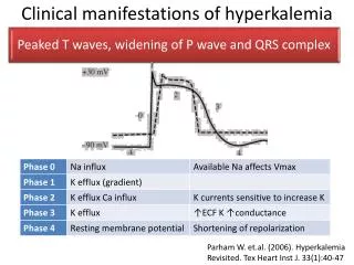
ECG changes — Tall peaked T waves with a shortened QT interval are usually the first findings • As the hyperkalemia gets more severe, there is progressive lengthening of the PR interval and QRS duration, the P wave may disappear, and ultimately the QRS widens further to a sine wave pattern. • Ventricular standstill with a flat line on the ECG ensues with complete absence of electrical activity.
Hyperkalemia: T wave in hyperkalemia is typically tall and narrow, but does not have to be tall (may be just narrow and peaked pulling ST segment). Tall T means > 2 big boxes in the precordial leads or >1 small box in limb leads, or T wave taller than QRS.
The progression and severity of ECG changes do not correlate well with the serum potassium concentration as illustrated by the following observations: • Rare patients have a normal ECG despite a serum potassium above 9.0 meq/L • ECG manifestations are more likely with rapid onset hyperkalemia, and the presence of concomitant hypocalcemia, acidemia, and/or hyponatremia
Given the unreliable sensitivity, serial measurements of the serum potassium concentration should guide therapy in stable patients with hyperkalemia. • The ECG cannot be reliably used to monitor the efficacy of hyperkalemia therapy • In addition, peaked T waves alone are not specific for hyperkalemia, being seen in the early phase of acute myocardial infarction and with early repolarization, and some patients with left ventricular hypertrophy
Conduction abnormalities and arrhythmias — Hyperkalemia can lead to a variety of conduction abnormalities and arrhythmias: • Conduction abnormalities that may be seen include right bundle branch block, left bundle branch block, bifascicular block, and advanced atrioventricular block • Cardiac arrhythmias associated with hyperkalemia include sinus bradycardia, sinus arrest, slow idioventricular rhythms, ventricular tachycardia, ventricular fibrillation, and asystole
PATIENT ASSESSMENT — Careful monitoring of the ECG and muscle strength are indicated to assess the functional consequences of hyperkalemia. • Severe muscle weakness and/or marked electrocardiographic changes, including conduction abnormalities and arrhythmias, are potentially life-threatening and require immediate treatment. • These manifestations usually occur when the serum potassium concentration is ≥7.0 meq/L with chronic hyperkalemia or possibly at lower levels with an acute rise in serum potassium.
خانم 60 ساله ي ديابتي به دليل فشا رخون بالا مراجعه مي كند. در معاينه نكته ي مثبتي ندارد BP= 160/90 دارد و در آزمايشات انجام شده BUN:20Cr:1.1Na:137 K: 6.1 بيمار تحت در مان با لوزارتان و آملوديپين مي باشد.. اقدام در ماني شما براي هايپر كالمي چيست؟ • قطع لوزارتان و استفاده از داروي ديگر آنتي هايپرتنسيو • استفاده از پودر كي اگزالات • قطع لوزارتان و استفاده از داروي ديگر آنتي هايپرتنسيو+ استفاده از پودر كي اگزالات • نياز به اقدام خاصي ندارد.
مرد 62 ساله اي با نارسايي كليه ي مزمن و كراتينين 2.1 mg /dL و پتاسيم نرمال به دليل فشا رخون بالا روي رژيم كم نمك قرار مي گيرد. دو هفته بعد متوجه ضعف عضلاني مي شود. در معاينه ي فيزيكي مختصر كاهش تورگور پوست و ضعف عضلات پروگزيمال يافته مي شود . ECG بيمار موج T بلند و پهن شدن موج P و كمپلكس QRS رانشان مي دهد. آزمايش هاي وي بصورت زير مي باشد: