Download
1 / 36
450 likes | 705 Views
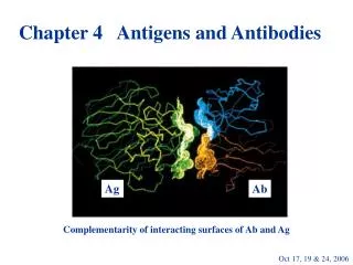
Basic structure of antibodies (Ab’s): Antibodies are heterodimers Chemical and enzymatic methods revealed basic Ab structure. Basic structure of antibodies (immunoglobulins): Tiselius and Kabat, 1939 immunized rabbits with ovalbumin (Ag) bled rabbits; this antiserum was

E N D
Basic structure of antibodies (Ab’s): Antibodies are heterodimers Chemical and enzymatic methods revealed basic Ab structure Ch. 4b
Basic structure of antibodies (immunoglobulins): Tiselius and Kabat, 1939 immunized rabbits with ovalbumin (Ag) bled rabbits; this antiserum was electrophoresed some antiserum was first incubated with ovalbumin and then electrophoresed; anti-ovalbumin Ab’s were “absorbed” from this serum Ch. 4b
p. 84 Ch. 4b
p. 85 Ch. 4b
p. 86 Ch. 4b
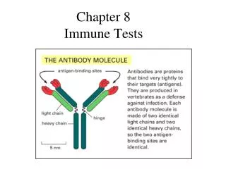
Enzyme digests IgG + papain 2 Fab + Fc IgG + pepsin 1 F(ab’)2 + small peptides Reduction and alkylation IgG 2 H chains + 2 L chains Light chain sequences revealed constant and variable regions There are 5 major classes of H chains and 2 types of L chains Ch. 4b
p. 87 Ch. 4b
Immunoglobulins (Ig) have multiple domains based on the Ig fold 4 (or 5) in heavy chain, 2 in light chain. Both heavy and light chains have 1 variable domain at the N-terminus about 110 amino acids in each domain Ig-fold: beta-pleated sheet intrachain disulfide bonds domains separated by “switch” region Ch. 4b
p. 88 Ch. 4b
p. 88 Ch. 4b
How are chains held together? disulfide bonds noncovalent interactions CDR’s (complementarity-determining regions) in variable domains bind Ag CDR’s also called hypervariable (hv) regions Rest of domain is called “framework” Ch. 4b
p. 91 Ch. 4b
p. 91 Ch. 4b
Constant-region domains CH1 and CL stabilize V regions contribute to antibody diversity Hinge flexibility Fab and Fc can move around it present in IgG, IgA, IgD IgE and IgM have no hinge, instead a fourth C domain Ch. 4b
CH2 has conserved glycosylation sites (some Ig subclasses have additional sites) Carbohydrate is sequestered between domains “Spreads out” the CH2; these regions tend to be biologically active Ch. 4b
Ch. 4b p. 88
Carboxy-terminal domain (CH3 or CH4) Can be membrane-bound or secreted Secreted form has hydrophilic tail Membrane-bound has hydrophilic spacer transmembrane sequence and cytoplasmic tail Ch. 4b
B cells express different classes of mIg at different developmental stages Immature B cell: mIgM only Mature B cell that has not seen antigen: mIgM and mIgD Memory B cell: mIgM, mIgG, mIgA, or mIgE mIg’s expressed sequentially on a single cell have identical Ag specificity Ch. 4b
Ab-mediated effector functions: - Opsonization is promoted by Ab - Ab’s activate complement (C) - Antibody-mediated cell-mediated cytotoxicity (ADCC) kills cells - Some Ab’s can cross epithelieal cells by transcytosis (IgA) Ch. 4b
p. 97 Ch. 4b
p. 98 Ch. 4b
IgG1 and IgG3 are most active Fix complement Bind to Fc receptors on phagocytes opsonization ADCC IgG4 binds to Fc receptors; does not fix complement IgG2 fixes complement moderately; has low affinity for Fc rceptors Ch. 4b
IgM pentamer (or hexamer), so 10 antigen- binding sites produced in primary response Ch. 4b
IgA most common antibody in body- not serum, but in secretions. Monomer in serum, multimer elsewhere helps protect portals of entry in body main protective antibody in breast milk Ch. 4b
p. 99 Ch. 4b
IgE Very low concentration in serum Binds to Fc receptors on basophils and mast cells; induces hypersensitivity response Ch. 4b
p. 100 Ch. 4b
IgD Very low concentration in serum Function of sIgD is not known * * * * * * * * * * Table 4-2 (p. 96) summarizes properties and biological activities of human serum Ig’s. Opsonization; C activation; ADCC; Transcytosis (e.g., Ab to mucosal surfaces, IgG across placenta - an example of passive immunity) Ch. 4b
p. 101 Ch. 4b
p. 102 Ch. 4b
The immunoglobulin superfamily Many proteins have a domain-like structure similar to immunoglobulins These other proteins do not share function and do not bind antigen What is the significance of this common structure? Ch. 4b
p. 103 Ch. 4b
p. 104 Ch. 4b
p. 105 Ch. 4b
Summary of antibody features Basic structure: two identical heavy chains, two identical light chains Antigen-binding and effector functions Membrane-bound and secreted forms Five heavy-chain isotypes that vary in function, serum concentration and serum stability (p. 96) Ch. 4b