Download

1 / 36
400 likes | 595 Views
Motor and Sensory Examination Dr. Bandar Al Jafen , MD Consultant Neurologist. MOTOR SYSTEM: GENERAL. There are five pattern of muscular weakness

E N D
Motor and Sensory Examination Dr. Bandar Al Jafen, MD Consultant Neurologist
MOTOR SYSTEM: GENERAL • There are five pattern of muscular weakness • Upper motor neurone (UMN) - increased tone, increased reflexes, pyramidal pattern of weakness (weak extensors in the arm, weak flexors in the leg. • Lower motor neuronr (LMN) – wasting fasciculation, decreased tone and absent reflexes. • Muscle disease – wasting, decreased tone, impaired or absent reflexes. • Neurmuscular junction – fatiguable weakness, normal or decreased tone, normal reflexes. • Functional weakness – normal tone, normal reflexes without wasting with erratic power.
WHAT TO DO Look at the position of the patient overall Look especially for a hemiplegic positioning, flexion of elbow and wrist with extension of knee and ankle. Look for wasting Look for fasciculation Test for tone Test muscle groups in a systematic way for power Test reflexes
TONE WHAT TO DO Ensure the patient is relaxed, or at least distracted by conversation. Repeat each movement at different speeds. Arms Take the hand as if to shake it and hold the forearm. First pronate and supinate the forearm. Then roll the hand round at the wrist. Hold the forearm and the elbow and move the arm through the full range of flexion and extension at the elbow. Legs Tone at the knee Put your hand behind the knee and lift it rapidly. Watch the heel. Hold the knee and ankle. Flex and extend the knee. Tone at the ankle Hold the ankle and flex and dorsiflex the foot.
Roll the knee Roll the wrist • WHAT YOU FIND • Normal: Slight resistance through whole range of movements. Heel will lift minimally off the bed. • Decreased tone: Loss of resistance through movement. Heel does not lift off bed when knee is lifted quickly. • Marked loss of tone = flaccid. • Increased tone: • - Resistance increases suddenly ('the catch') heel easily leaves bed when knee is lifted quickly – spasticity. • - Increased through whole range, as if bending a lead pipe – lead pipe rigidity. Regular intermittent break in tone through whole range – cogwheel rigidity. • - Patient apparently opposes your attempts to move his limb – Gegenhalten or paratonia.
Special situations • Myotonia – slow relaxation following action. • Dystonia – patient maintains posture at extreme or movement with contraction of agonist and antagonist. • Precussion myotonia may be demonstrated when a muscle dimples following percussion with a patella hammer. Most commonly sought in abductor pollicis brevis and the tongue. • WHAT IT MEANS • Flaccidity or reduced tone – common causes: lower motor nurone or cerebellar lesion; rare causes: myopathies, 'spinal shock' (e.g. early after a stroke), chorea. • Spasticity: upper motor lesion. • Rigidity and cogwheel rigidity: extrapyramidal syndromes – common causes: Parkinson's disease, phenothiazines. • Myotonia (rare) – cause: myotonic dystrophy (associated with frontal balding, ptosis, cataracts and cardiac conduction defects) and myotonica congenita. Percussion myotonia may be found in both conditions.
MOTOR SYSTEM: ARMS Testing elbow flexion Testing shoulder abduction
Testing elbow extension Testing finger extension Testing finger abduction Testing finger flexion
Testing finger abduction Testing thumb abduction
Testing strength of supraspinatus Testing strength of brachioradialis
MOTOR SYSTEM: LEGS Testing hib extension Testing hip flexion
Testing knee extension Testing knee flexion Testing plantarflexion of the foot Testing dorsiflexion of the foot
Testing inversion of the foot Testing eversion of the foot
MOTOR SYSTEM: REFLEXS Reflexes can be graded: 0 = absent + = present only wit reinforcement 1+ = present but depressed 2+ = normal 3+ = increased 4+ = clonus Testing the supinator reflex Testing the biceps reflex
Testing the triceps reflex Testing the knee reflex
Reinforcement Reinforcement If any reflex is unobtainable directly ask the patient to perform a reinforcement manoeuvre. In the arms ask the patient to clench his teeth as you swing the hammer. In the legs ask the patient either to make a fist, or to link hands across his chest and pull one against the other, as you swing the hammer. Demonstration of clonus At the ankle: Dorsiflex the ankle briskly, maintain the foot in that position, a rhythmic contraction may be found. More than three beat is abnormal. At the knee: With the leg straight take the patella and bring it briskly downwards; a rhythmic contraction may be noted. Always abnormal. The ankle reflex – three ways to get it
WHAT YOU FIND AND WHAT IT MEANS • Increased reflex or clonus – this indicates upper motor neurone lesion above the root at that level. • Absent reflexes: • - generalized – indicates peripheral neuropathy • isolated – indicates either a peripheral nerve or, more commonly, a root lesion. • Reduced reflexes (more difficult to judge) – occurs in a peripheral neuropathy, muscle disease and cerebellar syndrome. • Pendular reflex– this is usually best seen in the knee jerk where the reflex continues to swing for several beats. This is associated with cerebellar disease. • Slow relaxing reflex– this is especially seen at the ankle reflex and may be difficult to note. It is associated with hypothyroidism.
WHAT IT MEANS • Babinski's sign positive- indicates upper motor neurone lesion. • Babinski's sign negative– normal. • No response– may occur with profound upper motor neurone weakness (toe unable to extend); may occur if there is a sensory abnormality interfering with the afferent part of the reflex.
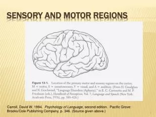
SENSATION: GENERAL There are five modalities of sensation. Sensory loss in the hand: a. Medium (red and ulnar (black) nerves Sensory loss in the hand: b. redial nerve Spinal cord section showing sensory input (red) from and motor output (black) to the right (R) side
Sensory loss in the arm: C. Axillary nerve Sensory loss in the leg: a. Lateral cutaneous nerve of the thigh; b. Common peroneal nerve; C. Femoral nerve d. Sciatic nerve Dermatomes in the arm
WHAT TO DO Vibration sense Use a 128 Hz tuning fork, those of higher frequency (256 or 512 Hz) are not adequate. Test– ask the patient to close his eyes, place the tuning fork on the bony prominence, ask if he can feel the vibration. Check– Check the patient reports feeling the vibration and not just the contact of the tuning fork. Strike the tuning fork and stop it vibrating immediately and repeat the test. If the patient reports that he feels vibration, demonstrate the test again. Joint position sense With the patient's eyes open show him what you are going to do. Hold the distal phalanx between your two fingers. Ensuring that your fingers are at 90o to the intended direction of movement, move the digit, illustrating which is up and which is down. Potential sites for testing vibration sense How to test joint position sense
Pin prick • Test – ask the patient to close his eyes then apply randomly sharp and blunt stimuli and note the patient's response. • Light touch • Use a piece of cotton wool. • Test – ask the patient to close his eyes, test the areas as for pin prick, apply the stimulus at random intervals. • Check – this is done by noting the timing of the response to the irregular stimuli. Frequently a pause of 10-20 seconds may be useful. • Special situation • Sacral sensation – this is not usually screened. It is essential to test sacral sensation in any patient with: • Urinary or bowel symptoms • Bilateral leg weakness • Sensory loss in both legs • Or where a cord conus medullaris or cauda equine lesion is considered. • Temperature sensation • Screening • It is usually adequate to ask a patient if the tuning fork feels cold when applied to the feet and hands. • Formal testing • Fill a tube with warm water and cold water.
Other modalities Two-point discrimination This requires a two-point discriminator – a device like a blunted pair of compasses. Test – gradually reduce distance between prongs, touching either with one or two prongs. Note the setting at which the patient fails to distinguish one prong from two prongs. Check – random sequence of one or two prongs allows you to assess testing. - Normal: index finger < 5mm; little finger < 7 mm; hallux < 10 mm. N.B. Varies considerably according to skin thickness. Compare right with left.
WHAT YOU FIND • Patterns of sensory loss • Sensory deficits can be classified into eight levels of the nervous system: • Single nerve • Root or roots • Peripheral nerve • Spinal cord • Brainstem • Thalamic sensory loss Glove and stocking loss Sensory loss associated with spinal cord lesions: a. Complete transverse lesion; b. Hemisection of the cord
f. Brainstem lesion; g. Thalamic sensory loss
CO-ORDINATION WHAT TO DO Test the gait In all tests compare right with left. Expect the right hand to be slightly better (in a right-handed person). Arms Finger-nose test Legs Heel – shin test The finger –nose test The heel-shin test
WHAT IT MEANS • Unilateral inco-ordination– ipsilateral cerebellar syndrome. • Bilateral inco-ordination– bilateral cerebellar syndrome. • Truncal ataxia, gait ataxia, without limb inco-ordination– midline cerebellar syndrome. • Unilateral cerellar syndrome– common causes: demyelination, vascular disease; rare causes: trauma, tumour or abscess. • Bilateral cerebellar syndrome– common causes: drugs (anti-convulsants), alcohol, demyelination, vascular disease; rare causes: hereditary cerebellar degenerations, paraneoplastic disorders, hypothyroidism. • Midline cerebellar syndrome: lesion of the cerebellar vermis – causes as for bilateral cerebellar syndrome.
GAIT • Always examine patient's gait. It is a co-ordinated action requiring integration of sensory and motor functions. The gait may be the only abnormality on examination. The most commonly seen are: hemiplegic, parkinsonian, marche a' petits pas, ataxic and unsteady gaits. • Romberg's test is conveniently performed after examination the gait. This is a simple test primarily of joint position sense. • Ask the patient to walk • Ensure you are able to see the arms and legs adeguately. • Is the gait symmetrical? • Gait can usually be divided into symmetrical and asymmetrical even though the symmetry is not perfect. • If symmetrical: • Look at the size of paces • Small or normal? • If small paces: • Look at the posture and arm swing • Stooped with reduced armswing – parkinsonian(may be difficult to start and stop –festinant). • Upright with marked armswing – marche a' petits pas.
If normal paces: • Look at the lateral distance between the feet • normal • widely separated - broad based • Legs unco-ordinated – cerebellar • Crossing over, toes dragged – scissoring. • Look at the knees • normal • knees lifted high – high-stepping. • Look at the pelvis and shoulders • normal • marked rotation of pelvis and shoulder –waddling. • Look at the whole movement • normal • disjointed as if forgotten how to walk, patient frequently appears rooted to pot – apraxic. • bizarre, elaborate and inconsistent – functional. • If asymmetrical • Is the patient in pain? • yes – painful or antalgic gait. • Look for a bony deformity • orthopaedic gait.
Does one leg swing out to the side? • yes – hemiplegic gait. • Look at the knee heights • normal • one knee lifts higher – foot drop. • Ask the patient to walk as if on a tight – rope (demonstrate) • if patient fall consistently – unsteady • may fall predominantly to one side. • Ask the patient to walk on his heels (demonstrate) • If unable to – foot drop. • Ask the patient to walk on his toes (demonstrate) • If unable - weakness of gastrocnemius. • Parkinsonian: indicates basal ganglion dysfunction – common causes: Parkinson's disease, major tranquillisers. • Scissoring: indicates spastic paraparesis – common causes: cerebral palsy, multiple scelrosis, cord compression. • Sensory ataxia: indicates loss of joint position sense (Romberg's positive) – common causes: peripheral neuropathy, posterior column loss (see below). • Cerebellar ataxia: veers towards side of lesion – common causes: drugs (e.g. phenytoin), alcohol, multiple sclerosis, cerebrovascular disease.
Waddling gait: indicates weak or ineffective proximal muscles – common causes: proximal myopathies, bilateral cogenital dislocation of the hip. • Apraxic gait: indicates the cortical integration of the movement is abnormal, usually with frontal lobe pathology – common causes:normal pressure hydrocephalus, cerebrovascular disease. • Hemiplegic: unilateral upper motor neurone lesion – common causes: stroke, multiple sclerosis. • Foot drop: common causes – unilateral: common peroneal palsy, pyramidal lesion, L5 radiculopathy. Bilateral: peripheral neuropathy. • Non-neurological gaits • Painful gait:common causes:arthritis, trauma – usually obvious. • Orthopaedic gait: common causes: shortened limb, previous hip surgery, trauma.
SUMMARY OF SCREENING NEUROLOGICAL EXAMINATION • If the history reveals no suggestion of focal neurological deficit, no speech disturbance and no disturbance of higher function, then you can use a screening neurological examination. • Screening neurological examination • Pupils – direct and consensual reactions. • Test fields to hand movements. • Fundoscopy. • Eye movements to pursuit on upgaze and lateral gaze. • Facial sensation to light touch with finger tip in all three divisions of trigeminal. • Facial movement – ‘screw up your eyes – show me your teeth’. • Mouth – ‘open your mouth’ (look at tongue) ‘and say “ahh” (observe palate). ‘Please put out your tongue’. • Test neck flexion. • Arms • - Look for wasting • - Test tone at wrist and elbow • - Observe outstretched arms with eyes closed (pronator test) • - Test power (shoulder abduction, elbow flexion and extension, finger extension and abduction and abductor pollicis brevis) • - Reflexes (biceps, triceps and supinator).
Legs • - Look for wasting • - Test tone at hip • - Test power (hip flexion and extension, knee flexion and extension, foot dorsiflexion and plantar flexion). • - Reflexes (knee, ankle and plantar response). • Sensation • - Test joint position sense in toes and fingers • - Test vibration sense on toes and fingers • - Test light touch and pinprick distally in hand and feet. • Coordination – test finger – nose and heel – shin. • Gait.