Download
1 / 54
540 likes | 542 Views
Neuronal Networks Laboratory. Sorting the connections with multi-electrode neuronal ensemble recording techniques: Single-unit and local field potential activity from rat to man Dr Rob Mason Institute of Neuroscience School of Biomedical Sciences

E N D
Neuronal Networks Laboratory Sorting the connections with multi-electrode neuronal ensemble recording techniques: Single-unit and local field potential activity from rat to man Dr Rob Mason Institute of Neuroscience School of Biomedical Sciences University of Nottingham Medical School and the LAB TEAM
Neuronal Networks Laboratory LAB TEAM epilepsyBen Coomber[Dr Mike O’Donoghue~Dept Neurology, QMC] Clare Roe schizophreniaDr Jill Suckling[Prof CA Marsden] Dr Dissanayake anxiety Dr Carl Stevenson[Prof CA Marsden] pain Dr Steve Elmes[Dr V Chapman] rodent USVs Beth Tunstall[Dr S Beckett] data analysisMargarita Zachariou[Prof S Coombes / Dr M Owen] [Mathematics Dept] Dr David Halliday (University of York) Prof D Auer (fMRI / phMRI ~ QMC)
Neuronal Networks Laboratory Sorting the connections with multi-electrode neuronal ensemble recording techniques To & From ~ rat prefrontal cortex • Seminar overview • Multichannel Electrophysiological Recording Technologies • Illustrate with reference to 2 experimental rodent model projects: • Hippocampus-mPFC ~ Epilepsy model – role of endocannabinoid system • Sensory gating in hippocampus & mPFC ~ Schizophrenia model – PCP effects • Amygdala-mPFC ~ Stress models –maternal separation & “drug stressors” • Left-Right mPFC~ • Rat language~ ultrasound vocalisations (USVs) & affective state
Sorting signal from noise Ensemble Neuronal Unit activity LFP activity DATA Analysis & Interpretation
Distribution of neurones contributing to signals recorded by tetrode array after Busaki 2004
Examples of Electrode Arrays 4-channel - independent manipulation Michigan MEAprobe 16-channels NBLabs 8-channel array Bionics 100-channel (“hedgehog”) array
Single-site recording • e.g. hippocampal sub regions (CA1 & CA3) • Multiple-site recording • e.g. prefrontal cortex & hippocampus MULTIPLE ELECTRODE ARRAY RECORDING in vivo • NBLabs • 16-channel micro-wire array • CA1 & CA3
Multiple Electrode Recordings in vivo • simultaneous multichannel spike & field potential recording • 64-channel MAP system • 32-channel MAP system – with CinePlex for behavioural studies • 64-chanel Recorder system • 16-chanel Recorder system • used MAP & Recorder for in vitro MEA studies with brain slices & neuronal cultures
100 0 Raw Signal (V) Units + EEG / LFP 100 Filtered Signal (V) Units 0 Impulse Events 0.20 TIME(s) 0.10 0 Extracellular Recording - signal filtering & discrimination • AP Spike discrimination:separate action potential (AP) signal from noise • AP amplitude detection / AP waveform shape recognition • Signal filtering: separate unit activity and Local Field Potentials (LFPs)
Electrode 1 Electrode 2 micro-electrodes nerve impulses (action potential “spikes”) Amplifier Neurones Multiple Neuronal (spike) Recording - two electrodes
Movie: Sorting of unit spike data using Principal Component Analysis • distinct AP spike waveforms represented as clusters in 3D space • 7 units isolated – each unit colour-coded
Multiple (ensemble) neurone recording • Advantages • Investigating neuronal ensemble/network function - closer to working “brain” • Good experimental design - fewer animals required (“3Rs” ~ Home Office) • Masses of data • Disadvantages • Masses of data • ? Data processing • ? Data interpretation
ENSEMBLE DATA - DISPLAY & ANALYSIS MASSIVE data sets Data Visualisation Emergent Properties Population Dynamics Unit activity Single units – spike rasters /FRH / ISIH / PSTHs burst analysis / Unit pairs – cross-correlation / coherence / Unit ensembles – PCA / ICA / synchrony index / PDC Local Field Potentials FFT / spectrograms LFP/EEG signal bands LFP-unit coherence LFP PDC
Neuronal Networks Laboratory • Dual/Triple site recordings ~ 64 channels simultaneous units & LFPs • VTA - mPFC • hippocampus - mPFC • amygdala - mPFC • mPFC – mPFC • spinal cord – thalamus – cerebral cortex • Systemic pharmacological manipulation • Local pharmacological manipulation • “injectrode” • integrated microiontophoresis with recording array • Electrical stimulation – periphery / CNS structures • Independent electrode array microdrive – 8 channel drive • Anaesthetised & awake-behaving preparations
Contralateral mPFC medial Pre-Frontal Cortex schizophrenia drugs of abuse VTA PVt Hippocampus nucleus Accumbens Amygdala SCN LAB EXPERIMENTAL DIRECTIONS mPFCx interconnectivity & functional context – in vivo studies bladder circadian - affective states • maternal separation • Depression • Stress • - Sensory Gating • - Schizophrenia • - Epilepsy USVs - affective states
Neuronal Networks Laboratory University of Nottingham Medical School Neuronal Networks Laboratory • Project #1: Epilepsy • kainate-induced epileptiform activity ~ TLE • functional network interactions ~ hippocampus mPFC • role of endocannabinoid system ~ CB1R pharmacology • perforant path stimulation-evoked seizure activity
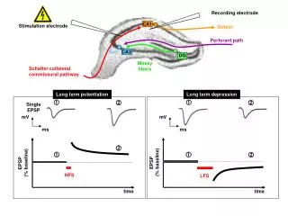
Endogenous cannabinoids (eCBs) identified e.g. anandamide (AEA) and 2-arachidonylglycerol (2-AG) Act at cannabinoid G protein coupled-Receptors: CB1 & CB2 - ? CB3 eCBs are synthesised post-synaptically on-demand. eCBs act at pre-synaptic CB1receptors. eCB reuptake occurs via a transporter. Metabolism: AEA by fatty acid amide hydrolase (FAAH) 2-AG by monoacylglycerol lipase (MGL). FAAH MGL Reuptake Endo-CANNABINOID System Figure taken from Wilson & Nicoll (2002) Science.
URB597 Metabolism by FAAH Anandamide Inhibits anandamide metabolism Epilepsy Study: AIMS • Kainic acid (KA): established convulsive agent producing seizures in awake rats ~ targeting temporal lobe Model uses KA, administered systemically (10mg/kg, i.p.) anaesthetized rats ~ ensemble neuronal unit and LFP activity • Study aims to establish whether URB597, selective inhibitor of FAAH enzyme (eCB levels), attenuates KA-evoked neuronal activity • Role of CB1 cannabinoid receptors assessed using selective CB1 antagonist AM251
Unit and LFP activity in mPFC and hippocampus Basal: Rat #1 Basal: Rat #2 KA + URB597: Rat #2 KA + Vehicle: Rat #1
Effects on hippocampal neural firing rate (~40 cells; n=5 rats) • Effects on spike-triggered averaging of mPFC LFPs Post-KA administration
Cross-correlation analysis ~ PFC – PFC / PFC- Hippocampal neuronal pairs # units n=2 n=12 Ref unit n=7
Cross-correlogram TF #1 Time-Series cross-correlograms – TF #1-13 correlation strength Time Frame #1-13 Time (s) +1s 0 -1s Time (s) correlation strength Time Frames #1-13 • Cross-correlation analysis of PFC neuronal pairs • Effect of kainate (10mg/kg; i.p.) administration at epoch 4
PDC Analysis – unit ensemble data • PDC was applied to identify the direction of activity between hippocampus • and mPFC - technique that has the potential to reveal the neuronal ensemble drives. Note: the magnitude of the classical coherence gives no information about directional connectivity - but its phase may do so.
Neuronal Networks Laboratory University of Nottingham Medical School Neuronal Networks Laboratory • Project #2: Schizophrenia • auditory-evoked sensory gating ~ hippocampus & mPFC • effects of PCP / ketamine~ model • effects of social isolation ~ model
Neuronal Networks Laboratory Sensory gating in hippocampus: A model for schizophrenia ? • Sensory gating: mechanism(s) by which irrelevant sensory information is filtered ~ enables efficient information processing • Auditory Conditioning-Test paradigm: measures reduction in auditory-evoked response produced by Test stimulus following a Conditioning stimulus • Stimuli: 3kHz sine-wave / 10ms duration / presented 500ms apart / 80-90dB • human P50 wave = rat N40 component • Gating absent in - schizophrenic patients (& family) • - normal volunteers given PCP / amphetamine • - rats given PCP / amphetamine
Units LFPs 1s LFP2 averaged 128 trials - 1 Trial # - 128 mV N40 N40 s Cs Ts Hippocampal CA3 - auditory-evoked unit & LFP activity event tone Averaged LFP T/C ratio = 55%
Single-unit PSTHs ‘ rasters histograms – “gating rats” CA3 region CA3 region dentate gyrus Spike raster PSTH Unit 1 Unit 2 Unit 3 Unit 4 LFP trial raster LFP
Hippocampal CA3 auditory-evoked unit & LFP activity Effects of PCP (1mg/kg i.p.) attenuates / abolishes sensory gating 1 Trail # 128 Basal [128 trials] T/C ratio = 32% i.e. exhibits gating 45mins after PCP T/C ratio = 66% gating attenuated
SUMMARY III Sensory Gating Studies • LFP studies: • Demonstrate sensory gating in isoflurane-anaesthetised rat : T/C ratio = 35 ± 15% • SG is abolished / attenuated following PCP : T/C ratio = 65 ± 5% • control ratsSG isunaffected by clozapine : T/C ratio 40% • Clozapine (5mg.kg-1) blocks action of PCP on SG : T/C ratio 35% • Unit studies: • Similar observations
Neuronal Networks Laboratory • Project #3: • Rat ultrasound vocalisations (USVs) & affective state • Sorting the pips from the squeaks • Role of nuc Accumbens in USV-mediated Behaviours ~50 kHz (Reward) call • (Brudzynski, 2001) • nucleus Accumbens - mPFCx functional connectivity
Neuronal Networks Laboratory • CURRENT APPROACHES & FUTURE DIRECTIONS • Human Studies • Neuro-Robotics & NeuroProsthetics • Hybrid Brain-Machine interfaces (HBMIs) • Cochlear implants • Monitoring & control of epileptic seizures • Robotic limbs
(A) Seizure control (B) Robotic arm control
Chips in the Brain ~ Brain-Machine Interfaces (BMIs) ~ Brain-Computer Interfaces Current issue (May 2007) Scientific American MIND 18 (2): 65-69
Brain-Computer Interfaces Signal choice & Algorithm Unit activity vs LFPs
Electrode array Implant Recording WorkStation – Plexon Inc
(1) ENSEMBLE RECORDINGS OF HUMAN SUBCORTICAL NEURONS AS A SOURCE OF MOTOR CONTROL SIGNALS FOR A BRAIN-MACHINE INTERFACE Parag G. Patil et al - Neurosurgery (2004) – Duke University ~ 32-channel PtIr 40m wire array ~ 4 Deep Brain Stimulation electrodes [Medtronics DBS] Unit recordings – 4 microwires Thalamic VOP/VIN STN [Plexon Inc MAP recording system]
Patient performance motor task STN recording ~ 24 units (2) ENSEMBLE RECORDINGS OF HUMAN SUBCORTICAL NEURONS AS A SOURCE OF MOTOR CONTROL SIGNALS FOR A BRAIN-MACHINE INTERFACE Parag G. Patil et al - Neurosurgery (2004) – Duke University
REFERENCES • Human / Primate Recording Reviews • LEARNING TO CONTROL A BRAIN–MACHINE INTERFACE FOR REACHING AND GRASPING BY PRIMATES • JM Carmena, et al • PLoS Biology ~ http://biology.plosjournals.org 1(2): 193-208 (2003) • ENSEMBLE RECORDINGS OF HUMAN SUBCORTICAL NEURONS AS A SOURCE OF MOTOR CONTROL SIGNALS FOR A BRAIN-MACHINE INTERFACE • PG Patil, JM. Carmena, Miguel AL Nicolelis & DA Turner • Neurosurgery 55(1): 27-38 (2004) • ASSISTIVE TECHNOLOGY & ROBITC CONTROL USING MOTOR CORTEX ENSEMBLE-BASED NEURAL INTERFACE SYSTEMS IN HUMANS WITH TETRAPLEGIA • JP Donoghue et al • J . Physiol 579(3) 603-611 (2007)
Neuronal Networks Laboratory University of Nottingham Medical School Neuronal Networks Laboratory That’s all folks Lab web site www.nottingham.ac.uk/neuronal-networks
in vivo Basal Bicuculline (7.5 mg.kg-1 i.v.) in vivo Basal Kainate (10 mg.kg- I i.v.)
Neuronal Networks Laboratory University of Nottingham Medical School Neuronal Networks Laboratory • Project #4: Affective state • Cortico-limibic network interactions • anxiety • effects of maternal separation • pharmacologically-induced (e.g. FG-7142) anxiety • behavioural sequalae • rodent ultrasound vocalisations
Neuronal Networks Laboratory University of Nottingham Medical School Neuronal Networks Laboratory • Project #5: • Nociception & pain management • dual spinal cord / supraspinal recording • Role of endocannabinoid system in normal physiology and pain (e.g. neuropathic) states
Mechanically-evoked response in somatosensory thalamus (VPM) noxious (65g) stimulation innocuous (7g) stimulation
Neuronal Networks Laboratory The Role of the CB2 Receptor in Nociceptive Processing: An in vivo electrophysiological study • Cannabinoid receptor agonists are antinociceptive. • CB1 predominantly expressed in the CNS but also present in the periphery. • CB2 agonists inhibit: • Acute pain [Zimmer et al.] • Inflammatory pain [Clayton et al.] • Neuropathic pain[Ibrahim et al.] • CB2 receptors located on: • Immune cells • Neuronal cells (?) [Griffin et al; Ross et al; Patel et al.] • CB2 agonists lack CNS side effects. • Development of potent selective CB2 ligands: • Agonist: JWH-133 Ki 3.4nM with a 200-fold selectivity over CB1 receptors. • Antagonist: SR144528 Ki 0.67nM with a 50-fold selectivity over CB1 receptors. • Aim: To determine the involvement of the CB2 receptor in nociceptive processing.
unit activity - nuc accumbens USV call start time Behavioural data Local Field Potential (LFP) USV Recorder input Spectrogram of specific calls [AviSoft] Combined unit / LFP with USV / behavioural recording
Recorded video Neuronal Networks Laboratory Neural data Behavioural Electrophysiology Circular arena recording using CinePlex Movie- rat HopScotch: 8-channel array in nuc. accumbens - 6 weeks post implant
DISCUSSION • Following KA, hippocampal units (~80%) show an increase in firing; while mPFC units show either a decrease (~80%) or increase (~20%) in firing rate. mPFC units lose their characteristic bursting pattern after KA administration. • CCH analysis shows that unit pair activity under basal conditions is more correlated within the mPFC compared to intra-hippocampal; mPFC appears to lead hippocampal firing. KA increased correlation within the hippocampus and mPFC; but the mPFC-hippocampal drive was lost. • PDC of unit population activity also shows basal mPFC-hippocampal directionality (predominantly at low frequencies); this initially decreases after KA administration, then later increases at all frequencies. • In basal conditions, PDC analysis of LFPs revealed evidence of information flow from CA3 to CA1 and reciprocal hippocampal-mPFC connectivity with predominant drive from mPFC to hippocampus. Following KA, there was increased drive from mPFC to hippocampus. • This alteration in functional connectivity in a seizure model has implications for memory and learning in epilepsy. • Caveat(s): • Need to consider possible influence of anaesthesia in directing “information flow” and/or (anaesthesia/KA-induced) short-term rewiring of neural circuitry. Other regions (not recorded) may be involved in communication between mPFC and hippocampus.