Download
1 / 38
400 likes | 1.62k Views
Plasmid DNA Isolation and Restriction Mapping. 生化科 林光輝. Plasmid DNA isolation Agarose gel electrophoresis Determine DNA fragment size; Restriction mapping. DNA. Contains all genetic information for that organism Size is usually expressed in base pairs (bp) 2 kinds of DNA:

E N D
Plasmid DNA isolation • Agarose gel electrophoresis • Determine DNA fragment size; Restriction mapping
DNA • Contains all genetic information for that organism • Size is usually expressed in base pairs (bp) • 2 kinds of DNA: • Chromosomal DNA • Makes up majority of DNA • Packed tightly into chromosomes • Plasmid DNA • Extrachromosomal DNA • Small circular molecules, usually 2-10 kb • Widely used in recombinant DNA technology
Isolation of DNA • A good prep should: • Not contain cellular proteins • Not contain RNA • Be of high molecular weight
The alkaline cell lysis method • Harvest bacterial cells • Resuspend in Tris-EDTA (RNase, lysozyme) • Add lysis buffer (NaOH+SDS) • Add sodium acetate PH6.5 • Centrifuge and discard the pellet • Further purification (special matrix)
Quality and Quantity of DNA • Spectrophotometer • Quantity: A260 = DNA 1 O.D. = 50 mg/ml DNA ___ O.D. x 50 mg/ml x 100 (dilution) = ___ mg/ml 1 O.D. • Quality:A280 = protein Purity = A260/A280 If ratio ≈ 2, then assume it is a good prep. If ratio > 2, then there is RNA contamination. If ratio < 1.6, then there is protein contamination.
Quality and Quantity of DNA • Gel electrophoresis • Quantity: Band intensity is semi-quantitative • Quality: Look for high MW DNA single clear band • Sheared DNA indicates poor quality smear
Basic Steps in Gene Cloning + + Host cell Transformed host cell Fragmented genomic DNA or cDNAs from chosen resource Recombinant DNA molecules Vector Amplification of recombinant molecule Host cell division 1 ‘gene’ purified in a clone Numerous cell divisions CLONE Colonies of transformed host cell clones growing on solid medium But how many individual clones needed to represent the entire genomic DNA or expressed genes of the resource?
Techniques of specific cleavage of DNA • Aim: to isolate and manipulate individual genes. • Means: Restriction endonucleases
What are Restriction endonucleases? • enzymes that attack and digest internal regions of the DNA of an invading bacteriophage but not that of the host. • First enzyme extracted from E. coli (cut randomly and not always close to the desired site).
Types of Restriction endonucleases • Type I and III: Possess both cutting (restriction) and protecting activity. • Protecting activity (modification by methylation). • Type I cuts at random sites.
Types of Restriction endonucleases • Type III cuts at specific sites quite near the recognition sequence. • but may be difficult to predict. • ATP required for source of energy.
Types of Restriction endonucleases Type II restriction enzymes are: • Invariably used in DNA science • They have several advantages: • 1) has only restriction activity; modification activity carried by a separate enzyme.
Types of Restriction endonucleases • 2) Each cuts in a predictable and consistent manner at a site within or adjacent to the recognition sequence. • 3) ATP not needed. Only a cofactor Mg++ is needed.
Types of Restriction endonucleases • Type II (continued). • Today, more than • 1200 type II have been isolated from a variety of prokaryotic organisms • More than 100 types are commercially available
Property of restriction enzymes • They break the phosphodiester bonds that link adjacent nucleotides in DNA molecules.
Nomenclature of Restriction endonucleases • 1) First letter: initial letter of the genus name of the organism from which the enzyme is isolated. • 2) Second and third letter: usually initial letters of the organism’s species name.
Nomenclature of Restriction endonucleases • 3) Fourth letter (if any): indicates a particular strain of organism • 4) Roman numerals: originally indicate the order in which enzymes from the same organism and strain are eluted from chromatography column.
Nomenclature of Restriction endonucleases More often, though, indicate the order of discovery.
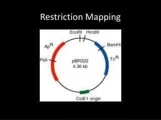
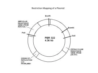
Examples of Type II restriction enzymes EcoRI E = genus Escherichia co = species coli R = strain RY13 I= first endonuclease isolated
Examples of Type II restriction enzymes BamHI B = genus Bacilus am = species amyloliquefaciens H = strain H I = first endonuclease isolated
Examples of Type II restriction enzymes HindIII H = genus Haemophilus in = species influenzae d = strain Rd III = third endonuclease isolated
Type II Restriction Enzymes Recognize symmetric DNA sequences = inverted repeats Most are 4-8 base pair long Cleave within recognition site 5’-GAATTC-3’ 3’-CTTAAG-5’ Only need Magnesium
Type II RE practical details 4, 5 or 6 bases 3 types of cuts - 5’ overhang, 3’ overhang, blunt This DNA continues on! 5’-GAATTC-3’ 3’-CTTAAG-5’ 5’-G-3’OH 5’P-AATTC-3’ 3’-CTTAA-P5’ HO3’-G-5’
Type II RE practical details 4, 5 or 6 bases 3 types of cuts - 5’ overhang, 3’ overhang, blunt This DNA continues on! 5’-GAATTC-3’ 3’-CTTAAG-5’ 5’-GAATT-3’OH 5’P-C-3’ 3’-C-P5’ HO3’-TTAAG-5’
Type II RE practical details 4, 5 or 6 bases 3 types of cuts - 5’ overhang, 3’ overhang, blunt This DNA continues on! 5’-GAATTC-3’ 3’-CTTAAG-5’ 5’-GAA-3’OH 5’P-TTC-3’ 3’-CTT-P5’ HO3’-AAG-5’
Sample Well 25 ng 1 kb ladder 0.8% Agarose Agarose Gel Electrophoresis Sybergold Detection Limit: ~0.2-0.5 ng DNA EtBr Detection Limit: ~5-10 ng DNA kb 12 10 8 6 4 2 1
Log (MW) is linearly related to mobility High % has no resolving power at high MW 10 kb 1 kb Low % has no Resolving power at low MW 100 bp
Star Activity (Relaxation of Specificity) . Under certain conditions, the enzymes are able to recognize and cleave nucleotide sequences which differ in some positions from the canonical site. For example, under extreme reaction conditions (i.e. low ionic strength), BamHI(recognition sequence GGATCC) is able to cleave the following sequences: NGATCC, GPuATCC and GGNTCC. This phenomenon has been called relaxed or star activity.
Star Activity In most practical applications of restriction endonucleases, star activity is not desirable. The analysis of a great number of reports on star activity suggest the following reasons for this phenomenon: • prolonged incubation time or a large excess of enzyme with respect to DNA; • high glycerol concentration (>5%) in the reaction mixture or the presence of other organic solvents, such as ethanol or dimethyl sulfoxide • low ionic strength or high pH values in the reaction buffer; • substitution of cofactor Mg2+ with other divalent cation (such as Mn2+ or Co2+).
Isoschizomers • Restriction endonucleases that recognize the same sequence are isoschizomers. Avi II and Fsp I TGC/GCA