Download
1 / 55
590 likes | 1.06k Views
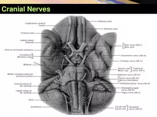
The cranial abdomen: Liver and Spleen. Tony Pease, DVM, MS Assistant Professor of Radiology North Carolina State University. Reading. Chapter 41 in Thrall. Imaging the cranial abdomen. Thickest part of the body Difficult to get enough contrast

E N D
The cranial abdomen:Liver and Spleen Tony Pease, DVM, MS Assistant Professor of Radiology North Carolina State University
Reading • Chapter 41 in Thrall
Imaging the cranial abdomen • Thickest part of the body • Difficult to get enough contrast • Place thickest part of the patient towards the cathode • Heel effect
Cranial abdomen • Multiple structures • Liver • Spleen • Stomach • Pancreas • Small intestine • Transverse colon
Other modalities MRI CT
Liver • Just caudal to the diaphragm • Cranial to the stomach • Liver size influences gastric axis
Liver or Spleen? LIVER! If it extends cranial to the antrum of the stomach Stomach
Hepatomegaly • Rounding of the liver margin • Extends well beyond the costal arch • Caudodorsal shift of the gastric axis
Causes for hepatomegaly • Hepatic venous congestion • Neoplasia (lymphoma) • Hyperadrenocorticism • Steroid hepatopathy • Diabetes mellitus • Hepatic lipidosis • Acute hepatitis
Radiographs • Since radiographs just give shape • Hard to narrow differentials • Need other modalities • Ultrasound • CT or MRI becoming more available
Focal hepatomegaly • Neoplasia • Abscess • Cyst • Biloma • Liver lobe torsion (rare)
Small liver • Hard to define • Upright gastric axis • Difficult to evaluate with ultrasound • Lungs get in the way
Differential diagnoses • Chronic liver disease • Cirrhosis • Hepatitis • Portosystemic shunt • Diaphragmatic hernia
Portosystemic shunt • Abnormal communication • Portal vein or tributary • Caudal vena cava or azygous vein • Multiple ways to detect
Good visualization Hepatic vasculature Invasive Surgical approach Contrast medium Complications Time Hypothermia Catheter removal Pros and Cons
Nuclear medicine • Gold standard • Administer radioisotope per rectum • 99m technetium pertechnetate • Enters colic vein to portal vein • Yes or no, but no anatomic data • Surgeons cannot use as a guide
Nothings perfect • Microvasculature dysplasia • No gross vessel problem • Defect is at the capillary level • Looks like normal scintigram
Ultrasound • Can be 95 – 100 % accurate • Is operator dependant • Patients usually quite small • Patients usually do not like process
Computed tomography • Still requires contrast medium • Usually debilitated patients • Less time with helical scanners • Can do 3D reconstruction
MRI • No contrast needed • Can do a “Time of Flight” • Allows blood to be its own contrast! • Moderate amount of time • Depends on magnet
Gall Bladder • Look for stones or mineral • Can see with radiography or ultrasound • Difficult to tell if important • Ultrasound can help • Especially with cholecystitis
Cholecystitis • Generally radiographs not helpful • Ultrasound helps more
The spleen • Generally surrounded by fat • Very clear on radiography • Sedation can increase spleen size
Spleen • Not many things happen to spleen • Separate into diffuse and focal disease • Radiographs give an idea • Ultrasound more helpful for seeing small lesions within parenchyma
Neoplasia Lymphoma Mast cell disease Congestion Sedation Right heart failure Splenic torsion IMHA Inflammation Infarction Nodular hyperplasia Extramedullary hematopoesis Diffuse spleen enlargement