Download
1 / 1
10 likes | 142 Views
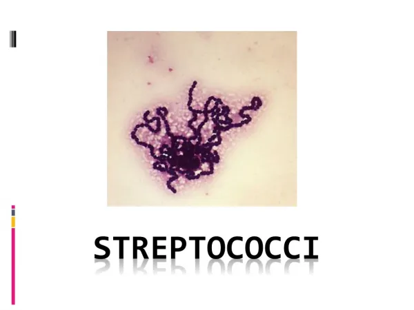
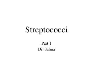
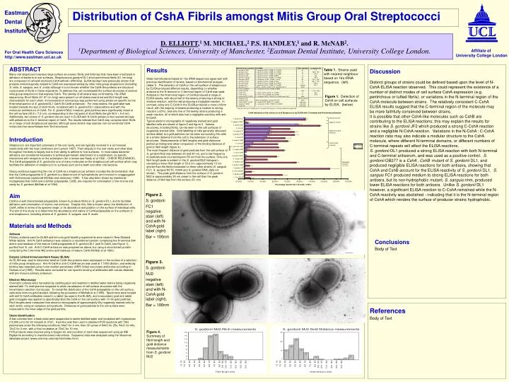
Figure 2. S. gordonii FC1 negative stain (left) and with N-CshA gold label (right) Bar = 100nm. Figure 3. S. gordonii MJ2 negative stain (left) and with N-CshA gold label (right). Bar = 100nm. Eastman Dental Institute.

E N D
Figure 2. S. gordonii FC1 negative stain (left) and with N-CshA gold label (right) Bar = 100nm Figure 3. S. gordonii MJ2 negative stain (left) and with N-CshA gold label (right). Bar = 100nm Eastman Dental Institute Distribution of CshA Fibrils amongst Mitis Group Oral Streptococci D. ELLIOTT,1 M. MICHAEL,2 P.S. HANDLEY,1 and R. McNAB2.1Department of Biological Sciences, University of Manchester, 2Eastman Dental Institute, University College London. Affiliate of University College London For Oral Health Care Sciences http://www.eastman.ucl.ac.uk ABSTRACT Many oral streptococci express large surface structures (fibrils and fimbriae) that have been implicated in adhesion of bacteria to oral surfaces. Streptococcus gordonii DL1 short peritrichous fibrils (61 nm long) are composed of cell-wall-anchored CshA adhesin (259 kDa). ELISA studies have previously shown that proteins immunologically related to CshA are expressed widely by other mitis group streptococci (including S. mitis, S. sanguis, and S. oralis) although it is not known whether the CshA-like proteins are structural components of fibrils in these organisms. To address this, we investigated the surface structures of several mitis group streptococci that express CshA. The identity of all strains was confirmed by 16s rDNA sequencing. Short fibrils (51-67 nm long) were present on all strains examined and fibril length was characteristic for each strain. Immunoelectron microscopy was performed using antiserum specific for the N-terminal portion of S. gordonii DL1 CshA (N-CshA antiserum). For most strains, the gold label was located towards the tips of short fibrils, consistent with S. gordonii DL1 observations and with the molecular architecture of CshA. For S. gordonii MJ2, however, gold particles were significantly closer to the cell surface (25.5 7.2 nm), corresponding to the mid-point of short fibrils (length 50.6 9.4 nm). Additionally, two strains of S. gordonii did not react in ELISA with N-CshA antiserum but reacted strongly with antiserum to the C-terminal region of CshA. The results indicate that CshA may comprise short fibrils on a range of oral streptococcal species, although some strains may express non-conventional CshA molecules that nevertheless form fibril structures. Introduction Streptococci are important colonisers of the oral cavity, and are typically involved in a commensal relationship with the host (Jenkinson and Lamont 1997). Their ubiquity in the oral cavity and other sites throughout the body is largely due to their ability to adhere to host surfaces. In most cases bacterial adhesins are considered necessary to achieve permanent attachment to a substratum, by specific interactions with receptors on the substratum (for a review see Hasty et al 1992 – CHECK RELEVANCE). The CshA polypeptide of S. gordonii is one of many molecules on the streptococcal cell surface which may facilitate the adhesion of streptococci to surfaces such as the teeth and other oral bacteria. Strong evidence supporting the role of CshA as a streptococcal adhesin includes the demonstration that that the CshA polypeptide of S. gordonii is a determinant of hydrophobicity and involved in coaggregation with Actinomyces naeslundii (McNab and Jenkinson 1992). It has also been shown by insertional mutagenesis that CshA and a similar polypeptide, CshB, are required for colonisation of the murine oral cavity by S. gordonii (McNab et al 1994). Aim CshA is a well characterised polypeptide, known to produce fibrils on S. gordonii DL1, and to facilitate adhesion and colonisation of murine oral surfaces. Despite this, little is known about the distribution of CshA, either in terms of its species range, or its abundance and position on the surface of individual cells. The aim of this study is to determine the abundance and nature of CshA polypeptide on the surfaces of oral streptococci, including strains of S. gordonii, S. sanguis, and S. oralis. Materials and Methods Antisera Primary antisera used for ELISA and immuno-gold labelling experiments were raised in New Zealand White rabbits. Anti-N-CshA antiserum was raised to a recombinant protein comprising the N-terminal 844 amino acid residues of the mature CshA polypeptide of S. gordonii DL1 (anti N-CshA, see Figure 1), purified from E. coli. Anti-C-CshA antiserum was prepared as above, but using a recombinant protein comprising the C-terminal 482 amino acid residues of mature CshA (McNab et al 1996). Enzyme Linked Immunosorbant Assay (ELISA) An ELISA was used to determine whether CshA-like proteins were expressed on the surface of a selection of mitis group streptococci. Anti-N-CshA or anti-C-CshA serum was used at 1:1000 dilution, and antibody binding was detected using horse raddish peroxidase (HRP) linked secondary antibodies according to Holmes et al (1995). Results were corrected for non-specific binding of antibodies with values obtained with pre-immune primary antiserum. Electron Microscopy Overnight cultures were harvested by centrifugation and washed in distilled water before being negatively stained with 1% methylamine tungstate to allow visualisation of cell surface structures with the transmission electron microscope. To reveal the distribution of the CshA polypeptide on the cell surface, cells were immuno-gold labelled, following the procedure of McNab et al (1999). Specimens were treated with anti N-CshA antibodies raised in a rabbit (as used in the ELISA), and a secondary goat-anti-rabbit gold-conjugate was applied to specifically label the CshA on the cell surface with 10 nm gold particles. Fibril lengths were measured from electron micrographs of approximately fifty negatively stained cells for each strain, using an eyepiece and graticule. Distances of gold particles to the cell surface were measured to the inner edge of the gold particle. Strain Identification A few colonies from a fresh plate were suspended in sterile distilled water and incubated with mutanolysin (10,000 u/ml) for 30 minutes at 37oC. A portion was then used in standard PCR reactions with TAQ polymerase under the following conditions; 94oC for 4 min, then 30 cycles of 94oC for 25s, 54oC for 40s, 72oC for 2 min, with a final incubation at 72oC for 10 min. PCR products were cleaned using a Qaigen kit, and a portion of each was sequenced using an ABI Bigdye kit according to manufacturers instructions. Sequence data was analysed using the ribosomal database project (www.cme.msu.edu/rdp/html/index.html). Results Strain identifications based on 16s rRNA sequences agree well with previous identification of strains, based on biochemical analysis (table 1). The detection of CshA on the cell surface of streptococci by ELISA produced different results, depending on whether antisera to the N-terminal or C-terminal region of CshA was used. Antisera to the N-terminal region of CshA produced a range of ELISA results, with about half of the strains producing a strong or medium reaction, and the rest producing a negligible reaction. In contrast, using anti-C-CshA in the ELISA produced a more uniform result, with the majority of strains producing a medium to strong reaction (>30%), and only five of the twenty strains producing a weak reaction, all of which also had a negligible reactivity with anti-N-CshA. Typical electron micrographs of negatively stained and gold labelled cells are shown in figure 2 and figure 3. Various structures, including fibrils, can be seen on the cell surfaces of negatively stained cells. Gold labelling of cells generally obscured surface detail, but gold particles can be seen surrounding the cells at a similar distance from the cell to the extension of surface structures. Measurements of fibril lengths and gold distances plotted as histograms allow comparison of the binding distance of gold to fibril length (figure 4). The most frequent distance of gold particles from the cell surface of S. gordonii MJ2 was between 20 and 30 nm, and a low frequency of particles were counted beyond 50 nm from the surface. Only one fibril length peak is evident in the S. gordonii MJ2 histogram, revealing a mean fibril length of 50.6 nm, approximately 15 nm shorter than the fibrils measured on three other strains (S. sanguis FC1, S. sanguis NCTC 10904 and S. gordonii PAR, data not shown). The peak gold distance from the surface of S. gordonii MJ2 is approximately 25 nm closer to the cell than the peak distance of fibril tips from the surface (51 nm) Table 1. Strains used with nearest neighbour based on 16s rRNA sequence. (left) Discussion Distinct groups of strains could be defined based upon the level of N-CshA ELISA reaction observed. This could represent the existence of a number of distinct modes of cell surface CshA expression (e.g. peritrichous or tufted fibrils), or variations in the N-terminal region of the CshA molecule between strains. The relatively consistent C-CshA ELISA results suggest that the C-terminal region of the molecule may be more faithfully conserved between strains. It is possible that other CshA-like molecules such as CshB are contributing to the ELISA reactions; this may explain the results for strains like S. gordonii JF2 which produced a strong C-CshA reaction and a negligible N-CshA reaction. Variations in the N-CshA : C-CshA reaction ratio may also indicate a modular structure to the CshA molecule, where different N-terminal domains, or different numbers of C-terminal repeats will affect the ELISA reactions. S. gordonii DL1 produced a strong ELISA reaction with both N-terminal and C-terminal antiserum, and was used as a positive control. S. gordonii OB277 is a CshA-, CshB-mutant of S. gordonii DL1, and produced negligible ELISA reactions for both antisera, showing that CshA and CshB account for the ELISA reactivity of S. gordonii DL1. S. sanguis FC1 produced medium to strong ELISA reactions for both antisera, but its non-hydrophobic mutant, S. sanguis nhm, produced lower ELISA reactions for both antisera. Unlike S. gordonii DL1 however, a significant ELISA reaction to C-CshA remained while the N-CshA reactivity was abolished - indicating that it is the N-terminal region of CshA which renders the surface of producer strains hydrophobic. Figure 1. Detection of CshA on cell surfaces by ELISA. (below) * Top panel= biotype I, middle panel = biotype II, bottom panel = biotype unknown/not applicable Conclusions Body of Text References Body of Text Figure 4. Summary of fibril length and gold distance measurements from S. gordonii MJ2