Download
1 / 60
600 likes | 1.07k Views
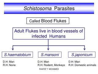
山东大学 医学院 病原生物学研究所 寄生虫学教研室 郭淑玲 . Schistosoma( 裂体吸虫 ). They belong to Genus Schistosoma, live in blood vessel and cause schistosomiasis. People call them blood flukes. There are four species infecting human body. They are:

E N D
山东大学 医学院 病原生物学研究所 寄生虫学教研室 郭淑玲
Schistosoma(裂体吸虫 ) They belong to Genus Schistosoma, live in blood vessel and cause schistosomiasis. People call them blood flukes. There are four species infecting human body. They are: 1. Schistosoma japonicum(日本血吸虫)is prevalent in Far East. In china, it is prevalent in Yangtze valley and south of Yangtze except Guizhou Province. The adults live in the portal vein system, causing liver cirrhosis(肝硬化)and portal vein hypertension syndrome(门脉高压征候群).
2. Schistosoma mekongi (湄公血吸虫) is merely distributed in Mekong River Valley(澜沧江:青海,西藏,云南――湄公河:老挝,泰国,柬埔寨), resembles Schitosoma japonicum except intermediate host. 3. Schistosoma haematobium(埃及血吸虫)widely spreads in Africa, chiefly in Nile River valley. The adults live in the vesical and pelvic plexus causing painless terminal haemuresis, renal failure complicated by the ureter obstruction. In the endemic area infection is so common that haemuresis is accepted as a sign of manhood in young boy. 4. Schistosoma mansoni(曼氏血吸虫)is distributed in Africa and focal area in Latin America. Lives in the portal and hemorrhoidal vein plexus, causing stool with fresh blood, liver cirrhosis and portal vein hypertension.
Schistosoma japonicum Only Schistosoma japonicum is found in China. Schistosomiasis is one of the “five major parasitic diseases”. It is prevalent in 13 provinces, city and autonomic regions (except Guizhou ) along Yangtze River Valley and south of Yangtze where are main areas for producing rice. I. Morphology 1. Adult worms are elongated cylindrical in shape, unlike other flukes. Two sexes are separate, gray white in color, but the female is much dark and slender, the male is shorter and thicker, sickle-like. In human body the male usually embraces the female into its gynecophoral canal (抱雌沟), appears “ K ”like (or the female usually resides in male’s gynecophoral canal).
Male: 10-20 x 0.5-0.55mm in size, oral sucker at top near by ventral sucker. Just behind the ventral sucker there is a longitudinal groove-gyncophoral canal in which the female normally resides. The esophagus is divided into two branches in front of the ventral sucker, and then unite to form a cecum at the posterior third part of the body. Seven testes are situated one by one, each has a delicate efferens which combine to form the vas deferens and dilate to become a seminal vesical opening in the genital pore just behind ventral sucker.
Female: Longer and slender than the male, much dark colored thread-like, 12-26x 0.1-0.3mm in size. The digestive system is similar to that of male. The vitellaria are located in the posterior part of the body surrounding the cecum. The unbranched, oval ovary lies in the mid-portion of the body. The uterus lies in the anterior portion of the body filled with 50-300 eggs arranged in a single row, arising from ootype to genital pore behind the ventral sucker.
Male and female schistosomes. (Drawn by Sylvia Treadgold) WHO
Paired male and female adult worms. The female schistosomulum is the darker, curled worm within the male's gynacophoric canal.
2. Mature egg is oval in shape, slight yellow in color, 89 x 67µ, shell is thin without an operculum but with a lateral spine. The content is a miracidium. Under the electron microscope there are many micro-tubules on the shell, through which the soluble egg antigen (SEA) secreted by a miracidium. 3. Cercaria is infective stage. It is composed of the body and forked tail (including tail stem and fork) and has 5 pairs of penetrating glands in the body.
oval in shape, slight yellow in color, 89 x 67 µ shell is thin without an operculum but with a lateral spine.
The small spine is generally not visible as the egg surface is often covered with facal debris.
oval in shape, slight yellow in color, 89 x 67 µ, shell is thin without an operculum but with a lateral spine.
II. Life cycle 1. Site of inhabitation: the portal vein system, mainly in the inferior mesenteric vein. 2. Infective stage: cercaria 3. Infective route: by skin 4. Intermediate hosts: Oncomelania snail (钉螺) 5. Reservoir hosts: mammals such as buffalo, cattle, wild rodents, goat, monkey, pig, fox. 6. Eggs are main pathogenic factor: (They are inlaid in the liver and intestinal wall. Some of them are discharged in feces to complete its life cycle). 7. The development in human body requires 25-30 days. Cercaria can live 1-3 days. Life span of the adults is about 20-30 years.
*8. Blood fluke is a special kind of flukes because of following characters: (1) The adult worms look like nematodes, elongated cylindrical in shape. (2) Two sexes are separate. (3) egg without operculum, but with a lateral spine. (4) only one intermediate host required. (5) The infective stage is cercaria. (6) The infective route is by skin. (7) The eggs are main pathogenic stage.
The infective inhabitation of Schistosoma japonicum: mesenteric vein
Diagram of the Life Cycle of Schistosoma Japonicum Liver Mesenteric vein Adults, ♂embosoms♀ 3 weeks migrate to in the portal vein system systemic circulation Lungs Liver cirrhosis Liver Eggs via Right heart IN HUMAN BODY intestinal 25-30days ulcers Blood stream Schistosomulum Discharged ( adolescent ) in feces cast off tail ────────────────────────────── Man and reservoir host Eggs get into water contact with contaminated In WATER 20-30℃ water, cercariae 6 hours penetrate the skin Miracidia hatch Carcariae invade escape from the snail Oncomelania snail Carcariae Daughter sporocysts Mother sporocysts
III. Pathology and Symptomatology 1.pathogenic mechanism of blood fluke
Eggs inlaid in the portal areas and intestinal wall SEA type IV allergy Local tissues necrosis, proliferation and fibrosis intestinal ulcers, Liver cirrhosis stool with blood, Pus and eggs Spleen Portal vein hypertension syndrome Enlargement Function failure Immunity Anemia Collateral circulation between portal vein and ascites, albumin, secondary infection vena cava are established emaciation Esophageal Umbilicus Hemorrhoid varicosity varicosity varicosity Hemorrhage of superior digestive tract Die of hepatic coma, superior digestive tract bleeding and infective complication
(1) Due to the cercaria and schistosomulum ( adolescent ): When human schistosome cercaria repeatedly penetrate the human skin, typeI allergy takes place. The cercarial dermatitis appears , petechiae(瘀点) and rashes ensue. The migration of the adolescents may induce localized pneumonitis and urticaria(荨麻疹). (2) Due to adults: The mechanical effect and toxic effect of adults and their metabolites cause mesenteric phlebitis, hepatitis, and abdominal pain; the immune complex may cause the damage to the kidney, schistosome nephritis results from typeIII allergy, the esinophile increase in peripheral blood.
* (3) Due to eggs: The most serious damage is done by eggs. Colon and liver are most seriously involved. 1) In liver: Soluble egg antigen Eosinophils infiltration Granuloma Eosinophilic abscess formation Fibrosis Liver cirrhosis (pipestem fibrosis) splenomegaly Portal vein hypertension ascites esophageal varicosity hemorrhoid varicosity varicosity varicosity surrounding the umbilicus
2) In intestine Soluble egg antigen Eosinophils infiltration Granuloma Eosinophilic abscess Ulceration Fibrosis or polyp
Intestinal schistosomiasis: eggs in the wall of the gut. WHO
2.Clinical manifestations (symptoms and signs ) (1) Initial phase: It is characterized by fever, dry cough (pneumonitis), urticaria. These phenomena are due to adolescents migration. *(2) Acute stage: The characteristics symptoms is dysentery. The patient may pass stool with blood, pus and mucus5-10 times per day, in which a large number of eggs can be found. Chill, fever, and malaise occur. *(3) Chronic stage:Chief manifestation of the patients are interval diarrhea or dysentery.The patients experience fatigue, general condition and strength deteriorate, loss of weight and interest, retardation of both physical and mental growth in children. Spleen and liver enlargement, anemia, in women menopause, sterility and abortion may occur. This stage may last from several years to 20 years.
*(5) Terminal stage is characterized by portal vein hypertension syndrome, common saying, abdomen distention looks like a big drum, emaciation looks like a fire wood (腹大如鼓,骨瘦如柴). Ascites, emaciation, varicosity, splenomegaly and anemia are commonly found. The patients die of secondary infection, upper digestive tract bleeding, hepatic coma. (6) Ectopic lesion: The damage to the central nervous system ( brain, spinal ) may cause paralysis (monoplegia, hemiplegia ).
abdomen distention looks like a big drum, emaciation looks like a fire wood. Ascites, emaciation, varicosity, and splenomegaly
abdomen distention looks like a big drum, emaciation looks like a fire wood.
IV. Dianosis The symptoms, signs and history of living in endemic areas only give a presumptive diagnosis. The definitive diagnosis depends on the pathogen examination. 1. Stool examination (1) Direct fecal smear for acute stage (2) Concentration method: Water sedimentation method and miracidia hatching test can be done at same time; nylon net method may be used. 2. Biopsy can be done by proctoscope for terminal stage. 3. Immunological tests are subsidiary for reference only.
V. Treatment and Prevention **1. Epidemiological investigation: Investigate whether Oncomelania snail can exist in local natural environment; local residents are used to defecate, work and play in the same water; and examine the pathogen: Examine the feces from local residents and domestic animals; also can dissect the suspicious reservoir hosts, such as buffalo, goat, wild rodents and etc. If the source of infection, intermediate hosts, transmitting route and susceptible crowd exist at same time and local, an endemic area of schistosomiasis can be confirmed.
2. Eliminate the source of infection (1) Treat the patients, carriers and domestic animals. Drug of choice for man: Praziqantel is pretty effective, side effects is very light. The other effective drugs , such as hexachloroparaxylol, bithionol may be used. (2) Kill the wild animals which may be infected. 3. Prevention (1) Health education is in progress, give up habits. (2) Control and deal with night soil. (3) Avoid directly contacting with the water contaminated by cercariae, lay up water in a container for 3 days, exposed to sun shine; put on protective clothes; apply some chemical repellent on the skin (dibutyl phthalate 邻苯二甲酸二丁酯 或灭蚴宁) (4) Kill the intermediate hosts and wild reservoir hosts. (5) Change the bad environment, realize modernization of agriculture.
VI. Epidemiology 1. Geographical distribution: The disease is prevalent in China, Japan, Philippines, Indonesia. In China, this disease is found in 13 province, city and autonomic region along the Yangtze River Valley and south of the Yangtze ( north from Jiangsu, Baoying county to south end Guangxi, Heng County ). In Taiwan, only animals, no humans are infected by S. japonicum. 2. Natural factors: (1) Endemic areas along river, stream, canal, irrigation canal with grass and vegetation growth.
(2) The geographil distribution of schistosmiasis is limited by the existence of Oncomelania snails. They live in warm ( temperature always over 0℃ ) and rainy ( average rainfall over 750mm per year ) regions where grass and vegetation grow along canals, streams and rivers with slowly flowing water or pools and lakes. (3) Source of infection include the patients, carriers and reservoir hosts. Rodents and buffaloes are especially dangerous sources of infection.
3. Social factors: • Habits: residents are used to defecate, work and play in the same water. The people who work on catching fish, planting rice, washing commodes, vegetables and clothes get infection easily. (2) Local economy and culture fall behind.