Download

1 / 16
200 likes | 568 Views
SCALP and MENINGES. Layers of the Scalp. S kin C onnective Tissue A poneurosis: Frontalis. Occipitalis. L oose Areolar Tissue P ericranium. Cutaneous Nerve Supply. CN V : (Trigeminal Nerve) Ophthalmic, maxillary, mandibular. Cervical spinal nerves: Ventral rami of C2, C3:

E N D
Layers of the Scalp • Skin • Connective Tissue • Aponeurosis: Frontalis. Occipitalis. • Loose Areolar Tissue • Pericranium
Cutaneous Nerve Supply • CN V: (Trigeminal Nerve) Ophthalmic, maxillary, mandibular. • Cervical spinal nerves: Ventral rami of C2, C3: Great auricular nerve (C2,3). Lesser occipital nerve (C2). • Dorsal ramus of C2: Greater occipital nerve.
Vascular Supply • Branches of external carotid: Occipital. Posterior auricular. Superficial temporal. • Branches of internal carotid: Supratrochlear. Supraorbital.
Meninges: Vascular Supply • Anterior meningeal arteries: From ethmoidal arteries and internal carotid. • Middle meningeal artery: From maxillary artery. • Emissary veins: Between dural venous sinuses and external veins. • Diploic veins. • Cerebral veins.
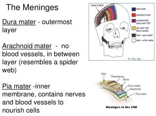
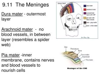
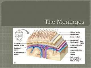
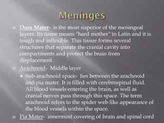
Meninges: Dura Mater • Tough outer connective tissue layer that forms sac around brain. • No epidural space in skull. • Encloses dural venous sinuses.
Meninges: Dura Mater • Reflections: Falx cerebri: Midline fold of dura mater extending between two cerebral hemispheres. Tentorium cerebelli: Dural fold located between cerebellum and occipital lobes of cerebral hemispheres.
Meninges: Dura Mater • Reflections: Falx cerebelli: Dural fold between two cerebellar hemispheres. Diaphragma sellae Dural fold over hypophyseal fossa.
Meninges: Dura Mater • Dural venous sinuses: Superior sagittal sinus: Lies along superior margin of falx cerebri. Inferior sagittal sinus: Lies along inferior margin of falx cerebri.
Meninges: Dura Mater • Dural venous sinuses: Straight sinus: Lies at intersection of falx cerebri and tentorium cerebelli. Confluence of sinuses: Common confluence of superior sagittal sinus and straight sinus.
Meninges: Dura Mater • Dural venous sinuses: Transverse: Begins at confluence of sinuses. Extends along edges of tentorium cerebelli. Right receives blood from superior sagittal sinus. Left receives blood from straight sinus.
Meninges: Dura Mater • Dural venous sinuses: Sigmoid: Continuation of straight sinus. “S”-shaped. Ends at jugular foramen: Joins internal jugular vein.
Meninges: Pia Mater • Innermost layer. • Closely applied to surface of brain. • Dips into fissures and sulci. • Forms sheath around blood vessels as they penetrate surface of brain.
Meninges: Arachnoid • Intermediate layer. • Attached to dura mater and pia mater: Separated from pia mater via: Subarachnoid space: Contains cerebrospinal fluid.
Meninges: Arachnoid • With many arachnoid villi: For reabsorption of cerebrospinal fluid. Arachnoid granulations may pit surrounding bone: Fovea granulares.
Meninges: Arachnoid • Subarachnoid cisterns: Choroid plexuses. Flow of CSF: Arachnoid villi. Arachnoid granulations. • Hydrocephalous: Obstructive. Communicating.