Download
1 / 1
10 likes | 209 Views
Eco. RV(7828). Eco. RV(5953). Eco. RV(6079). Eco. RV(2889). Eco. RV(3015). Eco. RV(1140). 35S. 35S. Kan-R. RB. RB. Kan-R. NOSpA. LB. LB. NOSpA. LBa1-primer. LBa1-primer. LBb1-primer. LBb1-primer. pROK2 T-DNA tandem. 8970 bp. Eco. RV(4). Eco. RV(5174). AP2C1. TAG.

E N D
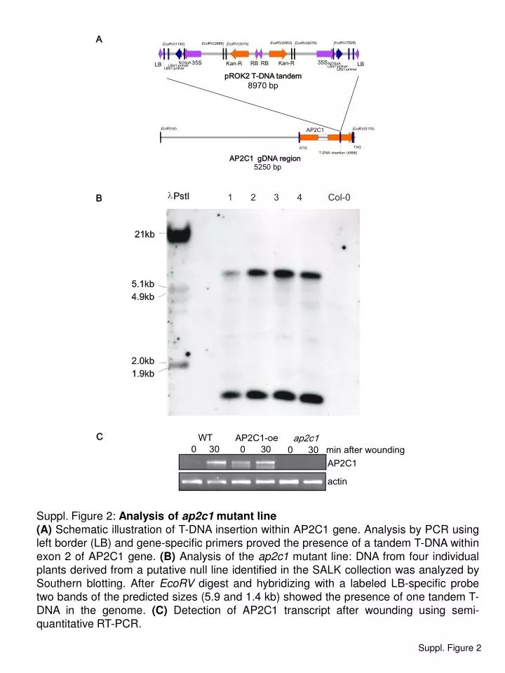
Eco RV(7828) Eco RV(5953) Eco RV(6079) Eco RV(2889) Eco RV(3015) Eco RV(1140) 35S 35S Kan-R RB RB Kan-R NOSpA LB LB NOSpA LBa1-primer LBa1-primer LBb1-primer LBb1-primer pROK2 T-DNA tandem 8970 bp Eco RV(4) Eco RV(5174) AP2C1 TAG ATG T-DNA insertion (4856) AP2C1 gDNA region 5250 bp l PstI PstI 1 2 3 4 Col-0 21kb 21kb 5.1kb 5.1kb 4.9kb 4.9kb 2.0kb 2.0kb 1.9kb 1.9kb WT AP2C1-oe ap2c1 0 30 0 30 0 30 min after wounding AP2C1 actin A B C Suppl. Figure 2: Analysis of ap2c1 mutant line (A) Schematic illustration of T-DNA insertion within AP2C1 gene. Analysis by PCR using left border (LB) and gene-specific primers proved the presence of a tandem T-DNA within exon 2 of AP2C1 gene. (B) Analysis of the ap2c1 mutant line: DNA from four individual plants derived from a putative null line identified in the SALK collection was analyzed by Southern blotting. After EcoRV digest and hybridizing with a labeled LB-specific probe two bands of the predicted sizes (5.9 and 1.4 kb) showed the presence of one tandem T-DNA in the genome. (C) Detection of AP2C1 transcript after wounding using semi-quantitative RT-PCR. Suppl. Figure 2