Download
1 / 13
130 likes | 270 Views
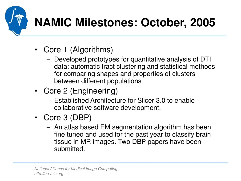
NAMIC Milestones: October, 2005. Core 1 (Algorithms) Developed prototypes for quantitative analysis of DTI data: automatic tract clustering and statistical methods for comparing shapes and properties of clusters between different populations Core 2 (Engineering)

E N D
NAMIC Milestones: October, 2005 • Core 1 (Algorithms) • Developed prototypes for quantitative analysis of DTI data: automatic tract clustering and statistical methods for comparing shapes and properties of clusters between different populations • Core 2 (Engineering) • Established Architecture for Slicer 3.0 to enable collaborative software development. • Core 3 (DBP) • An atlas based EM segmentation algorithm has been fine tuned and used for the past year to classify brain tissue in MR images. Two DBP papers have been submitted.
Algorithms: Diffusion Tensor Image Processing • DTI images capture information about local isotropic nature of neural structure • Ideally should differentiate strongly oriented structures (e.g. axons) from more diffuse structures • Scientific challenge is to compare shapes and other properties of white matter tracts across populations
Algorithms: Challenges • To compare tracts: • Need algorithms to extract from DTI images • Need algorithms to cluster into anatomically coherent collections • Need algorithms to measure properties along clusters • Need statistical methods for comparing shapes and properties of clusters between different populations • Have completed initial prototypes of all these algorithmic requirements
Algorithms: Impact • Clustering and statistical comparison tools will allow neuroscientists: • To identify and spatially isolate structural and functional differences (at the level of specific areas of the brain) between normal subjects and patients • To identify specific changes in white matter tract properties in correlation with other structural changes (e.g. coherence of local orientation) • To track changes in local structure of white matter tracts as disease progresses
Example of clustering across population Lauren O’Donnell, C-F Westin
Anterior Inferior Right Left Example identifying abnormality in cluster (due to tumor) Lauren O’Donnell, C-F Westin, Alex Golby
Example of change in fractional anisotropy due to MS lesion We observed a significant drop in FA (Fractional Anisotropy) value of the tracts where the lesion was located. Mahnaz Maddah, Simon Warfield, Daniel Goldberg
Core 2: Slicer 3.0 Architecture Established • The Engineering Core, working from requirements established by Core 1 and Core 3 have established an architecture for the next generation Slicer. • This architecture protects the algorithm investment of Core 1 while providing flexibility in the interfaces presented to the Core 3 DBP’s. • The software architecture isolates algorithms from the user interfaces. This will permit the same software to be run in batch, pipelined with LONI, executed on the UCSD Grid and support the interactive interfaces for the DBP’s. • In addition, the Slicer 3.0 architecture will support other software that conforms the the architecture command interface. This may include Matlab programs and other non-NA-MIC software. Bill Lorensen, Steve Pieper
Slicer 3.0 Architecture User Desktop Slicer3.0 Algorithms ITK VTK Slicer Modules Scripts of Slicer Mods VTK Apps Using ITK Batch Programs Non-NAMIC Cmd tools LONI Pipeline Birn Grid Data/Compute Bill Lorensen, Steve Pieper
Core 3: DBP Harvard Collaboration with MIT for Segmentation • Design of an atlas based EM segmentation algorithm developed by K. Pohl (MIT) which was then fine tuned for the classification of brain tissue in MR images. • Harvard group has been using this technique for about a year now and two DBP papers have been submitted: One on the effect of Schizotypal Personality Disorder on Neocortical Gray Matter and the other is on Neocortical Gray Matter Volume in First Episode Schizophrenia and First Episode Affective Psychosis. Sylvain Bouix, Martha Shenton
Atlas based EM Segmentation • Gray-scale image of a female with schizotypal personality disorder. • The same image with automatically segmented tissue types overlaid. CSF is color-coded by turquoise, gray matter by red-brown and white matter by pink. • The same image with neocortex and ventricle extracted by manual exclusion of non-neocortical structures from the automatically segmented image. On the right, CSF is color-coded by green, gray matter by brown, and white matter by yellow on right; left color codes as in part B. The lateral ventricle is color-coded by dark blue on the right and dark green on the left. The ROI definition illustrated in part C was used for the analysis. Sylvain Bouix, Martha Shenton
Segmentation References • Smaller Neocortical Gray Matter and Larger Sulcal CSF Volumes in Neuroleptic-Naive Females with Schizotypal Personality Disorder. Min-Seong Koo, Chandlee C. Dickey, Martha E. Shenton, Na Young Ji, Sylvain Bouix, Kilian M. Pohl, James J. Levitt, Motoaki Nakamura, Robert W. McCarley. Submitted to Arch. Gen Psychiatry • Neocortical Gray Matter Volume in First Episode Schizophrenia and First Episode Affective Psychosis: A Cross-sectional and Longitudinal MRI Study. Motoaki Nakamura, Dean F. Salisbury, Yoshio Hirayasu, Sylvain Bouix, Kilian M. Pohl, Takeshi Yoshida, Min-Seong Koo, Martha E. Shenton, and Robert W. McCarley. In preparation for submission to Arch. Gen Psychiatry. • Anatomical Guided Segmentation with Non-Stationary Tissue Class Distributions in an Expectation-Maximization Framework. Kilian M. Pohl , Sylvain Bouix, Ron Kikinis, W. Eric L. Grimson. 2004 IEEE International Symposium on Biomedical Imaging, Arlington VA., April 15-18, 2004, pp. 81 – 849
DTI References • White Matter Tract Clustering and Correspondence in Populations. Lauren O'Donnell, Carl-Fredrik Westin. Accepted to MICCAI 2005 • A method for clustering white matter fiber tracts. O’Donnell L, Kubicki M, Shenton ME, Dreusicke MH, Grimson WEL, Westin CF. AJNR (In Revision). • DTI and MTR Abnormalities in Schizophrenia: Analysis of White Matter Integrity. M. Kubicki, H.-J. Park, C.-F. Westin, P. Nestor, R. Mulkern, S. E. Maier, M. Niznikiewicz, E. Connor, J. Levitt, M. Frumin, R. Kikinis, F. A. Jolesz, R. McCarley, M. E. Shenton. Neuroimage 2005. • Fronto Temporal Disconnectivity in Schizotypal Personality Disorder: A Diffusion Tensor Imaging Study. M. Nakamura, R. W. McCarley, M. Kubicki, C. C. Dickey, M. A. Niznikiewicz, M. M. Voglmaier, L. J. Seidman, S. E. Maier, C.-F. Westin, R. Kikinis, M. E. Shenton. Biological Psychiatry 2005.