Download
1 / 28
380 likes | 729 Views
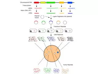
Is there an insert?. A.f. insert. Each organism has a specific set of restriction enzymes. Eco RI from E scherichia co li Bam HI from B acillus am yloliqueraciens Pvu I and Pvu II are different enzymes from same strain. Originally purified by individual labs, Nathans, Smith

E N D
Is there an insert? A.f. insert
Each organism has a specific set of restriction enzymes • EcoRI from Escherichia coli • BamHI from Bacillus amyloliqueraciens • PvuI and PvuII are different enzymes from same strain. • Originally purified by individual labs, Nathans, Smith • Now supplied by companies - GE, NEB, Promega, BRL Ch. 3-1
Restriction enzymes - endonucleases, Cleave a specific DNA sequence Protect bacteria from phage infection, digest phage DNA after infection Cellular DNA protected by methylases - block restriction enzyme activity Ch. 3-1
Restriction enzymes are used for cloning and analyzing DNA fragments
Sequence Recognition and cleavage: a) 5' overhang EcoRI GAATTC G pAATTC CTTAAG CTTAAp G b) 3' overhang KpnI GGTACC GGTAC pC CCATGG Cp CATGG c) Blunt end SmaI CCCGGG CCC pGGG GGGCCC GGGp CCC Ch. 3-2
Sequence Recognition and cleavage: d) Degenerate: AvaII GGWCC: GGTCC, GGACC AvaI CPyCGPuG CTCGAG Py stands for pyrimidine- T or CCTCGGG Pu stands for purine - A or GCCCGAG CCCGGG C TCGAG C CCGGG CCCGAG GAGCT C GGGCC C GAGCCC DdeI CTNAG: CTAAG, CTGAG, CTCAG, CTTAG BbsI cleaves GAAGACNN CTTCTGNNNNNN Ch. 3-2
Sequence Recognition and cleavage: e) Isoschizomers:Different enzymes cut the same seq. MboI, Sau3A, DpnII GATC N pGATCN CTAG NCTAGp N f) Overlaps: Two enzymes that give the same overhang BamHI GGATCC G pGATCC CCTAGG CCTAGp G Sau3ANGATCN N pGATCN NCTAGN NCTAGp N Ch. 3-2
Activity: in units which corresponds to a specified level of enzyme activity. NEB defines a unit as: “One unit of restriction endonuclease activity is defined as the amount of enzyme required to completely digest 1 g of substrate DNA in a total reaction volume of 0.05 ml in one hour using the NEB buffer provided.” Ch. 3-3
Restriction enzymes are proteins with optimal conditions Activity of an enzyme can change under different conditions: pH- 7.5, 8.0, 8.5 salt concentration- 20 mM, -150 mM divalent cations- Mg++ reducing reagent- DTT carrier protein-BSA temperature- 37C, RT, 60C • Before setting up a restriction digest check to make sure that you are using the proper conditions! Ch. 3-4
| A | B | C | D | E | F | H | K | M | N | P | R | S | T | X |Z | Enzyme Supplied NEBuffer % Activity in NEBuffers 1 2 3 4 Aat II 4 0 50 50 100 Acc I 4 50 50 10 100 Acc65 I 3 + BSA 10 75 100 25 Aci I 3 25 50 100 50 Acl I 4 + BSA 10 10 0 100 Acu I 2 + SAM 50 100 50 100 Afe I * SE-Y 25 50 25 100 Afl II 2 + BSA 50 100 25 100 Afl III 3 + BSA 25 75 100 50 Age I 1 100 50 10 75 Ahd I 4 + BSA 25 75 0 100 Ale I 4 10 20 10 100 Alu I 2 100 100 75 100 Alw I 4 50 100 10 100 AlwN I 4 50 100 50 100 Apa I @25°C 4 + BSA 25 50 0 100 ApaL I 4 + BSA 100 100 10 100 ApeK I @75°C 3 25 75 100 50 Apo I @50°C 3 + BSA 10 75 100 75 Asc I 4 0 10 10 100 Ase I 3 NR 75 dd100 NR AsiS I 3 + BSA 50 100 100 50 Ava I 4 10 75 10 100 Ava II 4 50 75 10 100 Avr II 2 100 100 50 100 Bae I @25°C 2 + BSA + SAM 50 100 50 75 BamH I Udd + BSA 75 100 50 75 Ban I 4 50 100 50 100 1 2 3 4 NEB Buffer Compatibility Chart NEBuffer 1 (yellow): 10mM Tris Propane (pH 7.0), 10 mM MgCl2, 1mM DTT NEBuffer 2 (blue): 10mM Tris (pH 7.9), 10 mM MgCl2, 50 mM NaCl, 1mM DTT NEBuffer 3 (red): 50mM Tris (pH 7.9), 10 mM MgCl2, 100 mM NaCl, 1mM DTT NEBuffer 4 (blue): 20mM Tris-acetate (pH 7.9), 10 mM Mg acetate, 50 mM K acetate, 1mM DTT
Setting up a restriction digest 1. Think about the experiment: Mapping or cloning? Determine which enzymes to use Are they in the freezer?? Determine buffer and reaction conditions Are the enzymes compatible? Ch. 3-4
Determine controls: uncut DNA parent vectors, size standards 3. Calculate how much DNA to add. Analytical or preparative? Ch. 3-5
Set up the Reaction: • Add in the following order: • Single Multiple • Sterile ddH2O 7.0 l 35.0 l • 10 X restriction buffer 2.0 l 10.0 l • Miniprep DNA (0.5 g) 10.0 l **none** • Enzyme (20 U/l) 1.0 l 4.0 l • Total volume 20.0 l 10.0 l Mix aliquot • 10.0 l DNA • The two most important rules in enzymes • Always keep enzymes on ice or in a cooler. • Always use a fresh tip when pipeting from the enzyme stocks. Ch. 3-6
5. Incubate reactions at the appropriate temperature for the appropriate time. Usually 37˚C and incubate 1 hr or more. 6. If running on a gel: Add gel loading dye EDTA - Stops reaction. Dyes (BPB and XC) - to help see sample while loading and monitor electrophoresis Glycerol - so sample sits at bottom of the well Ch. 3-6
DNA Amplification by PCR Ch. 3-5 Ch. 3-9
SP CTCCGAGATCTGGACGAGC> CTCCGAGATCTGGACGAGCTTTTTTTTTTTTTCTCGGGAAGCGCGCCATT 1 ---------+---------+---------+---------+---------+ 50 GAGGCTCTAGACCTGCTCGAAAAAAAAAAAAAGAGCCCTTCGCGCGGTAA INSERT | KpnI SmaI EcoRI PstI BamHI | | | | | GTGTTGGTACCCGGGAATTCGGCCATTATGGCCTGCAGGATCCGGCCGCC 61 ---------+---------+---------+---------+---------+ 100 CACAACCATGGGCCCTTAAGCCGGTAATACCGGACGTCCTAGGCCGGCGG XbaI XhoI HindIII | | | TCGGCCCAGTCGACTCTAGACTCGAGCAAGCTTATGCATGCGGCCGCAAT 121 ---------+---------+---------+---------+---------+ 180 AGCCGGGTCAGCTGAGATCTGAGCTCGTTCGAATACGTACGCCGGCGTTA TCGAGCTCACTTGGCCAATTCGCCCTATAGTGAGTCGTATTACAAT 181 ---------+---------+---------+---------+------ 196 AGCTCGAGTGAACCGGTTAAGCGGGATATCACTCAGCATAATGTTA <GCGGGATATCACTCAGCATAATGTTA- BP Ch. 3-5
1. Dilute aliquots of your miniprep plasmid DNA samples 50-fold. Label four fresh microfuge tubes and combine 98 l of H2O with 2 l of plasmid DNA. Mix each sample tube by vortexing. Lab 6-4
Prepare the following 5 Rxns. PCR Mix: • 1 Rxn.5 Rxns. PCR Mix • Sterile ddH2O 18.0 l 90 l • SP Primer (10 pmole/l) 2.5 l 12.5 l • BP Primer (10 pmole/l) 2.5 l 12.5 l • Diluted Vector (~2 ng) 2.0 l** (DO NOT ADD DNA!) • =25.0 l Lab 6-4
3. Obtain a strip of 4 Tubes of “PCR Beads.” • Label tubes • To each tube add: • one bead • 23 l of the “5 Rxn. PCR Mix. • 2 l of the appropriate diluted DNA • Mix each by gently tapping the tube. Lab 6-4
I Initial Denaturation 94oC for 5 minutes (Complete denaturation) II Amplification (Repeats steps 30X) 94oC for 1 minute (Denaturation of target DNA) 50oC for 1 minute (Annealing of primer to template DNA) 72oC for 1 minute (Elongation to produce new DNA strand) III Additional Elongation 72oC for 5 minutes (Insures all DNA strands are full length) IV Soak 4oC for ON. (Helps samples be stable)
Agarose Gel Electrophoresis: DNA is negatively charged Electric field causes the DNA fragments migrate through the gel to the positive electrode Smaller fragments migrate through the gel faster Separates DNA fragments on the basis of size Ch. 3-12


![[INSERT SPEAKER’S NAME] [INSERT TITLE] [INSERT DATE]](https://cdn3.slideserve.com/6899353/slide1-dt.jpg)