Download
1 / 32
320 likes | 456 Views
Research X-ray Safety Fundamentals. Office of Research Safety 114 Long Hall. Goal of this Training:. Define x-rays Explain the hazards of x-ray devices used in research List the requirements and responsibilities for the safe use of x-ray devices

E N D
Research X-ray Safety Fundamentals Office of Research Safety 114 Long Hall
Goal of this Training: • Define x-rays • Explain the hazards of x-ray devices used in research • List the requirements and responsibilities for the safe use of x-ray devices • Help you recognize and respond to unsafe conditions
What is Radiation? Radiation is energy in the form of waves or particles. Radiation of a high enough energy is called : IONIZING RADIATION Ionizing radiation includes x-rays, gamma-rays, beta particles, alpha particles, and neutrons. Without the use of monitoring equipment, radiation is undetectable. Humans are not able to see, feel, taste, smell, or hear ionizing radiation.
What are X-rays? • X-rays are a form of electromagnetic radiation produced when electrons are deflected from their original paths or change their orbital levels around the atomic nucleus. X-rays are capable of traveling long distances through air and most other materials. Like gamma rays, x-rays require more shielding to reduce their intensity than do beta or alpha particles. • X and gamma rays differ primarily in their origin: • x-rays originate in the electronic shell • gamma rays originate in the nucleus
X-rays X-rays were discovered in 1895when Wilhelm Conrad Roentgen observed that a screen coated with a barium salt fluoresced when placed near a cathode ray tube. Roentgen concluded that a form of penetrating radiation was being emitted by the cathode ray tube and called the unknown radiation: X-rays
X-ray Tube An x-ray tube requires a source of electrons, a means to accelerate the electrons, and a target to stop the high-speed electrons.
X-ray Interactions • In passing through matter, energy is transferred from the x-ray photon to electrons and nuclei in the target material. An electron can be ejected from the atom with the subsequent creation of an ion. The amount of energy lost to the electron is dependent on the energy of the incident photon and the type of material through which it travels. • There are three basic methods in which x-rays interact with matter: • photoelectric effect • Compton scattering • pair production
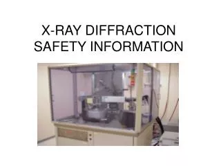
Analytical X-rays • Two main uses: • Diffraction [XRD] • X-ray scattering from crystalline materials. “fingerprint” of crystalline atomic structure. Check known library vs. unknown sample. • Fluorescence [XRF] • Analytical method for determining the elemental composition of a substance.
Hazards of Analytical X-ray Equipment The primary beam: The primary beam is most hazardous because of the extremely high exposure rates. Exposure rates of 4 x 10⁵ R/min at the port have been reported for ordinary diffraction tubes. Leakage or scatter of the primary beam through cracks in ill fitting or defective equipment: The leakage or scatter of the primary beam through apertures in ill fitting or defective equipment can produce very high intensity beams of possibly small and irregular cross section. Penetration of the primary beam through the tube housing, shutters or diffraction apparatus: The hazard resulting from penetration of the useful beam through shutters or the x-ray tube housing is slight in well designed equipment. Adequate shielding is easily attained at the energies commonly used for diffraction and florescence analysis. Diffracted rays: Diffracted beams also tend to be small and irregular in shape. They may be directed at almost any angle with respect to the main beam, and occasionally involve exposure rates of the order of 80 R/h for short periods.
Putting fingers in X-ray beam to change sample • Aligning X-ray beam visually • Modification of shielding • Failure to realize X-rays are emitted from several ports • Failure to read & follow manufacturers X-ray operating instructions • Any of these actions could cause an unnecessary exposure!
Safety Interlocks Safety interlocks may consist of switches or other devices that prevent operation of the x-ray device with shielding removed. X-ray devices should NEVER be operated with the safety interlocks bypassed. A written procedure must be approved by the Radiation Safety Officer PRIOR to performing any work with safety interlocks defeated.
Symptoms of Local X-ray Overexposure The most common effects from a large radiation exposure from an x-ray device is reddening of the skin (erythema). With a dose of a few hundred rem the superficial layers of the skin are damaged and the skin will redden in a fashion similar to a sunburn. The erythema effect will most often reverse itself within a few weeks. Contact the Radiation Safety Officer immediately if an overexposure is suspected! 656-7165
Diagnostic X-rays Two main types of diagnostic x-ray devices: Radiograph– a picture with film or image is sent direct to computer screen Fluoroscope – a real time “moving” inspection on inside functions Diagnostic radiology is the branch of medicine that has traditionally been known for taking and reading x-rays. Like every other field of medicine, technology has radically changed this specialty forever. Diagnostic radiology is the nucleus of almost every physician’s diagnosis. Being able to detect disease sooner and pinpoint its location more accurately is a huge factor in stopping disease in its tracks.
Industrial X-rays X-rays are used for non-destructive testing which has applications in a wide range of industries. Non-destructive testing (NDT) means the X-ray beam inspects the integrity of industrial products or processes without damaging the items under observation. The NDT field thatuses radiation is called Industrial radiography. Industrial X-ray machines are used primarily to find defects in castings, structures, and welds. These units can also help to find foreign material in food products. X-ray machines are used for the inspection of luggage at airports and buildings.
X-ray Effects The effects of x-ray exposure depends upon: • Duration - How fast the dose is delivered. • Energy - How much energy was in the x-ray • Low Energy (<50 KeV) - damage only to skin or outer part of body • High Energy - damage to internal organs • Total Dose - The magnitude of the dose
Unsafe Conditions • Examples of unsafe conditions: • Access door interlocks do not work, shielding has been damaged, or viewing window is cracked. • IF AN UNSAFE CONDITION ARISES WITH YOUR X-RAY DEVICE : • Stop work! • Turn power OFF to X-ray (An X-ray requires power to produce radiation) • Notify your Principal Investigator and the Radiation Safety Officer @ 656-7165
Radiation Protection: Time The dose of radiation a worker receives is directly proportional to the amount of time spent in a radiation field. Thus, reducing the time by one-half will reduce the radiation dose received by one-half. Operators should always work quickly and spend as little time as possible next to x-ray equipment while it is operating.
Radiation Protection: Distance Radiation exposure decreases rapidly as the distance between the worker and the X-ray device increases. The decrease in exposure from a point source, such as an X-ray tube, can be calculated by using the INVERSE SQUARE LAW: This law states that the amount of radiation at a given distance from a point source varies inversely with the square of the distance. For example, doubling the distance from an x-ray tube will reduce the dose to one-fourth of its original value. Maintaining a safe distance, therefore, represents one of the simplest and most effective methods for reducing radiation exposure to workers. Using the principle of distance is especially important when working around open beam analytical x-ray equipment.
Radiation Protection: Shielding Radiation exposure can also be reduced by placing an attenuating material between a worker and the x-ray tube. The energy of the incident x-ray photon is reduced by Compton and photoelectric interactions in the shielding material. Substances such as lead, that are very dense and have a high atomic number, are very practical shielding materials because of the abundance of atoms and electrons that can interact with the x-ray photon. Shielding is often incorporated into the equipment, such as the metal lining surrounding the x-ray tube. It may also consist of permanent barriers such as concrete and lead walls, leaded glass, and plastic movable screens in the case of analytical x-ray equipment.
Open Beam Devices This an OLD open beam x-ray diffraction device. New diffraction x-ray devices for research must be contained in an fully shielded – interlocked cabinet.
XRD Devices The x-ray tube, detector and sample are contained in housing that provides shielding to the user and others in lab. The access doors are interlocked and will shut off x-rays when opened. The large viewing area is made possible by using leaded glass or Plexiglas.
Diffraction cont. A small compact “totally enclosed” research X-ray device.
Radiography Table This is an x-ray tube in a collimated lead housing. The beam is pointed down to the table. The table is where the patient is placed and contains a slot for the film. This is the mobile shield for the operator. It is designed to protect the operator from scattered x-rays (primarily from patient). This is the control panel. The operator can select x-ray ON (exposure) time in fraction of minutes, the energy of x-ray (in kVp) and current applied (higher current = more x-rays).
Fluoroscopic C-arm When this “C-arm”x-ray device is used the operator and support staff MUST wear a lead apronand whole body dosimeter badge.
SC DHEC Regulations • X-ray devices must be registered with the South Carolina Department of Health, Bureau of Radiation Control. • Each x-ray system MUST meet state requirements. • Each system must be reviewed by the Radiation Safety Officer (RSO).
X-ray Requirements • If you acquire any x-ray devices you MUST notify the Radiation Safety Officer! • Radiation Safety inspects x-ray devices annually. • Each system must have a RSO approved radiation protection plan (RPP). • X-ray users must be approved by device Principal Investigator. • X-ray users need to complete Research X-ray Safety Trainingcourse prior to unsupervised use of an x-ray device.
Responsibilities of X-ray Owners & Users • Operate x-ray device only as specified in manufacturers operating instructions. • Notify Radiation Safety Officer of any repairs, modifications, disposal, or relocation of x-ray device.
Personnel and Area Monitoring Devices • All personnel who are likely to receive radiation exposure approaching 10% of the maximum occupational dose limit, and all users of industrial devices shall wear a personnel monitoring device. • Whole-body personnel monitoring devices will be worn routinely in the area of the breast pocket, collar, or waist. • Pregnant radiation workers shall wear a whole-body personnel monitoring device during the pregnancy. • Users of open-beam analytical x-ray equipment are required to wear a whole body and a ring badge • Finger ring TLD badges are required for maintenance activities of analytical x-ray equipment that requires presence of the primary beam when any local component in the system is adjusted, disassembled, or removed.
Pregnant Radiation Workers Pregnant radiation workers should: • Notify the Radiation Safety Officer as soon as her pregnancy is known (confidentiality will be maintained). • Limit her exposure to less than 500 mrem during the pregnancy. • Wear a whole-body personnel monitoring device during the pregnancy. • Keep her exposure to the very lowest practical level by reducing the amount of time spent in a radiation area, increasing the distance from a radiation source, and using shielding.
Personnel Monitoring Most analytical x-ray devices do not require users to be issued personnel monitoring devices. X-ray users should address any radiation safety concerns to the Radiation Safety Officer @ 650-7165.
Example of a Radiation Protection Plan (RPP) All personnel involved in using a University X-ray device must review this program and will be held accountable for violations. Any PI that may have a research need to purchase, borrow, or build a radiation generating device (X-ray) shall notify the Radiation Safety Officer (RSO). Radiation Safety will inspect x-ray devices and facilities annually. Any changes to an x-ray device (new tube, design modifications, etc.) MUST be approved by the RSO.This x-ray machine will be used as it is currently configured and approved for operation by the RSO. This machine will be operated in accordance with the manufacturers operating and safety procedures. A restricted area will be designated as needed by the RSO to protect personnel against undue risks from exposure to radiation. X-ray device users will be persons authorized by the Principal Investigator and/or RSO. Minors (age less than 18) or members of the general public are not allowed to operate x-ray device without prior approval of the RSO. Members of the public will be considered to be all persons other than those involved in the authorized use, surveillance, or inspection of this machine. Declared pregnant workers may use x-ray after a dosimeter is obtained from the RSO. The dosimeter device shall be worn at all times while using x-ray device.
This completes the training presentation. The link below will direct you to a short quiz to see how much knowledge you retained. If you fail to complete the quiz and registration page your training will not be recorded by Research Safety. http://www.clemson.edu/research/safety/training/xray/quiz.html If the link does not open please copy and paste the address into a web browser.