Download

1 / 45
460 likes | 954 Views
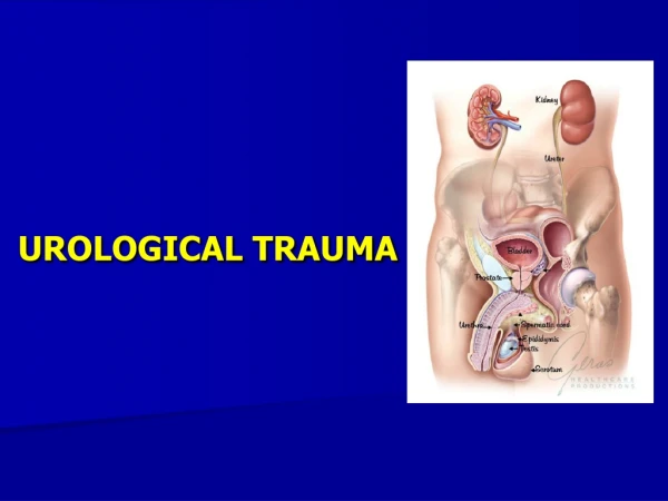
UROLOGICAL TRAUMA. RENAL TRAUMA. Background Renal trauma occurs in approximately 1-5% of all traumas. Renal injuries are the most common injuries of the urinary system.

E N D
RENAL TRAUMA Background • Renal trauma occurs in approximately 1-5% of all traumas. • Renal injuries are the most common injuries of the urinary system. • Blunt trauma directly to the abdomen, flank, or back is the most common mechanism, accounting for 80-85% of all renal injuries. • The kidney is well protected by heavy lumbar muscles, vertebral bodies, ribs, and the viscera anteriorly. • Kidneys with existing pathologic conditions such as hydronephrosis or malignant tumors are more readily ruptured from mild trauma.
Ethiology • Trauma may result from motor vehicle accidents, fights, falls, and contact sports. Vehicle collisions at high speed may result in major renal trauma from rapid deceleration and cause major vascular injury. • Fractured ribs and transverse vertebral processes may penetrate the renal parenchyma or vasculature. • Gun-shot and knife wounds cause most penetrating injuries to the kidney; any such wound in the flank area should be regarded as a cause of renal injury until proved otherwise. • Associated abdominal visceral injuries are present in 80% of renal penetrating wounds.
Classification • Minor renal trauma (85% of cases)- Renalcontusion or bruising of the parenchyma is the most common lesion. Subcapsular hematoma in association with contusion is also noted. Superficial cortical lacerations are also considered minor trauma. These injuries rarely require surgical exploration. • Major renal trauma (15% of cases)-Deep corticomedullary lacerations may extend into the collecting system, resulting in extravasation of urine into the perirenal space. Large retroperitoneal and perinephric hematomas often accompany these deep lacerations. Multiple lacerations may cause complete destruction of the kidney. Laceration of the renal pelvis without parenchymal laceration from blunt trauma is rare.
Classification • Vascular injury (about 1% of all blunt trauma cases) • Vascular injury of the renal pedicle is rare but may occur, usually from blunt trauma. • There may be total avulsion of the artery and vein or partial avulsion of the segmental branches of these vessels. • Vascular injuries are difficult to diagnose and result in total destruction of the kidney unless the diagnosis is made promptly.
Clinical Findings • The clinic of the closed damage of kidney depends on its degree. • Each kind of trauma is accompanied by characteristic manifestations and general signs, which are pain and intumescence in lumbar region, haematuria.
Clinical Findings • Symptoms: • Pain in lumbar region on the side of damage is observed in 80- 95 % of cases of isolated traumas of kidney and in 10-20 % of combined injuries. It is dull or acute with irradiation in inguinal region or external sexual organs. • Associated injuries such as ruptured abdominal viscera or multiple pelvic fractures also cause acute abdominal pain and may obscure the presence of renal injury. • Catheterization usually reveals hematuria. • Retroperitoneal bleeding may cause abdominal distention, ileus, nausea and vomiting.
Clinical Findings • Signs: • Initially, shock or signs of a large loss of blood from heavy retroperitoneal bleeding may be noted. Ecchymosis in the flank or upper quadrants of the abdomen is often noted. • Lower rib fractures are frequently found. • Diffuse abdominal tenderness may be found on palpation; an "acute abdomen" indicates free blood in the peritoneal cavity. • A palpable mass may represent a large retroperitoneal hematoma or perhaps urinary extravasation. If the retroperitoneum has been torn, free blood may be noted in the peritoneal cavity but no palpable mass will be evident. • The abdomen may be distended and bowel sounds absent.
Diagnostics • Diagnostics of the isolated closed renal damage is not difficult in general. • The anamneses, presence of trauma signs and hemorrhagies, pain in lumbar region, positive Pasternatsky’s symptom testify probability of renal trauma.
Diagnostics • Laboratory Findings: • Microscopic or gross hematuria is usually present. • The hematocrit may be normal initially, but a drop may be found when serial studies are done. This finding represents persistent retroperitoneal bleeding and development of a large retroperitoneal hematoma. • Persistent bleeding may necessitate operation.
Diagnostics: Chromocystoscopia • Chromocystoscopy, if possible, also helps to establish the correct diagnosis. • This method of research sometimes allows to find a location of bleeding (that it is very important in case of combined trauma), to analyse functions of damaged and opposite kidney, state of urinary bladder wall. • However for a choice of method of treatment it is necessary to know the character of damage and its localization.
X-Ray Findings • Observing radiography (KUB): the method allows to find damage of bones, to suspect presence of retroperitoneal hematoma (contours of kidney and lumbar muscles are absent). • Excretory urography gives an opportunity to define the side of damage, anatomical and function status of injured and opposite kidney.
X-Ray Findings • X-ray signs of renal damage are weak and later spreading of X-ray contrast solution in calyces-bowling systems, subcapsular and retrorenal spreading of X-ray contrast, deformation of renal bowl and calyces. .
X-Ray Findings • On angiogramms one can see violation of arterial and venous circulation attached to marginal injuries, filling of pararenal tissue with X-ray contrast due to injuries of renal artery branches.
Ultrasonography • Ultrasound scans can detect renal lacerations but cannot definitely assess their depth and extent. In addition, they do not provide functional information. • During the evaluation of blunt trauma patients, ultrasound scans were more sensitive and specific than intravenous pyelography (IVP) in minor renal trauma. • Another possible role for ultrasound may be for serially evaluating stable renal injuries for the resolution of urinomas and retroperitoneal haematomas.
Computed tomography (CT) • Staging begins with an abdominal CT scan, the most direct and effective means of staging renal injuries. • This noninvasive technique clearly defines parenchymal lacerations and urinary extravasation, shows the extent of the retroperitoneal hematoma, identifies nonviable tissue, and outlines injuries to surrounding organs such as the pancreas, spleen, liver, and bowel.
T R E A T M E N T • The treatment can be conservative and operational. • The majority of the experts choose tactic of expectation.
T R E A T M E N T • Bed regimen is provided within 10-20 day. • Measures to stop bleeding (administration of haemostatic agents, hemo- and plasmotransfusion), administration of analgetics, antibiotics of a wide spectrum of action, and also dynamical overseeing by arterial pressure. • Antibiotics are used for pyelonephritis prophylactics.
T R E A T M E N T • Indications to operative treatment: • а) internal bleedings in case of isolated renal damage, which are accompanied by an anaemia, decrease of arterial pressure, fast pulse; • b) hematuria within a day with worsening of general state of the patient; • c) hematoma in lumbar region, which is slowly growing; • d) combination of renal damage and organs of abdominal cavity or thorax.
TREATMENT • The operation should be maximum savings and directed on the decision of two tasks - stopping of bleeding and normalization of urine outflow.
INJURIES OF THE URINARY BLADDER • Bladder injuries occur most often from external force and are often associated with pelvic fractures. (About 15% of all pelvic fractures are associated with concomitant bladder or urethral injuries.) • Iatrogenic injury may result from gynecologic and other extensive pelvic procedures as well as from hernia repairs and transurethral operations.
Classification • closed and open • isolated and combined • intraperitoneal, retroperitoneal and mixed.
Clinical Findings • Symptoms: • There is usually a history of lower abdominal trauma. • Blunt injury is the usual cause. • Patients ordinarily are unable to urinate, but when spontaneous voiding occurs, gross hematuria is usually present. • Most patients complain of pelvic or lower abdominal pain.
Clinical Findings • Signs: • Heavy bleeding associated with pelvic fracture may result in hemorrhagic shock, usually from venous disruption of pelvic vessels. • An acute abdomen indicates intraperitoneal bladder rupture. • A palpable mass in the lower abdomen usually represents a large pelvic hematoma. • On rectal examination, landmarks may be indistinct because of a large pelvic hematoma.
Clinical Findings • Laboratory Findings: • Catheterization usually is required in patients with pelvic trauma but not if bloody urethral discharge is noted. • When catheterization is done, gross or, less commonly, microscopic hematuria is usually present. • Urine taken from the bladder at the initial catheterization should be cultured to determine whether infection is present.
X-Ray Findings • A plain abdominal film generally demonstrates pelvic fractures. There may be haziness over the lower abdomen from blood and urine extravasation. • An intravenous urogram should be obtained to establish whether kidney and ureteral injuries are present.
X-Ray Findings • Bladder disruption is shown on cystography. • Retrogradual cystography help to differentiate penetrating and unpenetrating, intraperitoneal and retroperitoneal ruptures of the bladder, locate urinary flow and approximate site of rupture. • The sign of retroperitoneal rupture is accumulation of X-ray contrast matter in perivesical fat tissue. • With intraperitoneal extravasation, free contrast medium is visualized in the abdomen, highlighting bowel loops.
Treatment • A.Emergency Measures: • Shock and hemorrhage should be treated. • B.Surgical Measures: • A lower midline abdominal incision should be made. As the bladder is approached in the midline, a pelvic hematoma, which is usually lateral, should be avoided. Entering the pelvic hematoma can result in increased bleeding from release of tamponade and in infection of the hematoma, with subsequent pelvic abscess. The bladder should be opened in the midline and carefully inspected. After repair, a suprapubic cystostomy tube is usually left in place to ensure complete urinary drainage and control of bleeding.
Treatment • In a case of retroperitoneal complete rupture of the bladder it is exposed by suprapubic extraperitoneal access carefully inspected and is juncture by two-row catgut junctures. • Drainage by means of epicystostomy is necessary. • Operation finish with drainage of perivesical and pelvic tissue. • In order to prevent formation of urinary flow, in all cases of retroperitoneal rupture of urinary bladder, perivesical space is drainage through obturatorial foramen or ischiorectal space.
Treatment • Intraperitoneal bladder ruptures should be repaired via a transperitoneal approach after careful transvesical inspection and closure of any other perforations. • The peritoneum must be closed carefully over the area of injury. • The bladder is then closed in separate layers by absorbable suture. • All extravasated fluid from the peritoneal cavity should be removed before closure. • At the time of closure, care should be taken that the suprapubic cystostomy is in the extraperitoneal position.
INJURIES Of THE URETHRA • Urethral injuries are uncommon and occur most often in men, usually associated with pelvic fractures or straddle-type falls. • Various parts of the urethra may be lacerated, transected, or contused. • Management varies according to the level of injury. • The urethra can be separated into 2 broad anatomic divisions: the posterior urethra, consisting of the prostatic and membranous portions, and the anterior urethra, consisting of the bulbous and pendulous portions
Clinical Findings • Symptoms: • Patients usually complain of lower abdominal pain and inability to urinate. A history of crushing injury to the pelvis is usually obtained. • Signs: • Blood at the urethral meatus is the single most important sign of urethral injury (Urethroragia). • Suprapubic tenderness and the presence of pelvic fracture are noted on physical examination. • A large developing pelvic hematoma may be palpated. • Perineal or suprapubic contusions are often noted. • Rectal examination may reveal a large pelvic hematoma with the prostate displaced superiorly.
Clinical Findings • Laboratory Findings: • Anemia due to hemorrhage may be noted. • Urine usually cannot be obtained initially, since the patient should not void and catheterization should not be attempted.
Clinical Findings • Instrumental Examination: • The only instrumentation involved should be for urethrography. • Catheterization or urethroscopy should not be done, because these procedures pose an increased risk of hematoma, infection, and further damage to partial urethral disruptions.
X-Ray Findings • Fractures of the bony pelvis are usually present. • A urethrogram (using 20-30 ml of watersoluble contrast material) shows the site of extravasation. • Ordinarily, there is free extravasation of contrast material into the perivesical space. • Incomplete prostatomembranous disruption is seen as minor extravasation, with a portion of contrast material passing into the prostatic urethra and bladder.
Treatment • Conservative therapy is effective for patients with recent nonpenetrating damage of urethra: rest, cool compresses, and antibiotics. • Within 7-8 days after trauma thermal procedures and resorption agents are prescribed. • In case of ischuria instead of a high cystotomy it is possible to perform troacar epicystostomy. • Shock and hemorrhage should be treated.
Treatment • Surgical Measures: • Urethral catheterization should be avoided. • Initialmanagement should consist of suprapubic cystostomy to provide urinary drainage. • A midline lower abdominal incision should be made, care being taken to avoid the large pelvic hematoma. • The bladder should be opened and carefully inspected for lacerations. If a laceration is present, the bladder should be closed with absorbable suture material and a cystostomy tube inserted for urinary drainage. • The suprapubic cystostomy is maintained in place for about 3 months. This allows resolution of the pelvic hematoma, and the prostate and bladder will slowly return to their anatomic positions.
Treatment • Urethral reconstruction -Reconstructionof the urethra after prostatic disruption can be undertaken within 3 months. • Before reconstruction, a combined cystogram and urethrogram should be done to determine the exact length of the resulting urethral stricture. • The preferred approach is a single-stage reconstruction of the urethral rupture defect with direct excision of the strictured area and anastomosis of the bulbous urethra directly to the apex of the prostate. • A 16F silicone urethral catheter should be left in place along with a suprapubic cystostomy. • Catheters are removed within a month, and the patient is then able to void
Complications • Stricture, impotence, and incontinence as complications of prostatomembranous disruption. • Stricture following primary repair and anastomosis occurs in about one-half of cases. If the preferred suprapubic cystostomy approach with delayed repair is used, the incidence of stricture can be reduced to about 5%. • The incidence of impotence after primary repair is 30-80% (mean, about 50%). Incontinence in primary reanastomosis is noted in one-third of patients. • Delayed reconstruction reduces the incidence to less than 5%.