Download
1 / 57
570 likes | 734 Views
Macromolecules of life structure And properties Aleksander L. Sieron Department General and molecular biology And genetics http://biolmolgen.slam.katowice.pl. Macromolecules of life structure And properties. Nucleic acids Proteins Sugars Lipids. Nucleic acids.

E N D
Macromolecules of life structure And properties Aleksander L. Sieron Department General and molecular biology And genetics http://biolmolgen.slam.katowice.pl
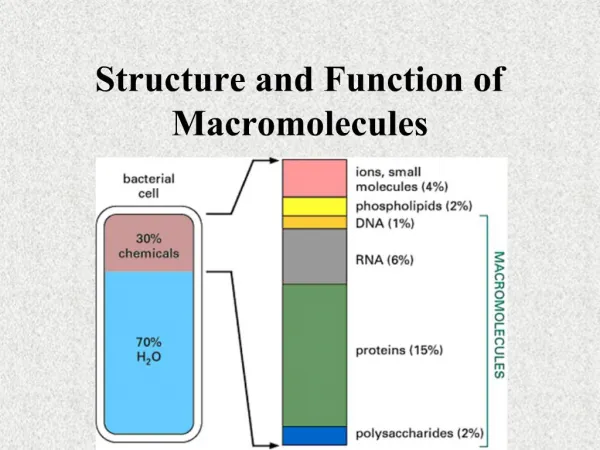
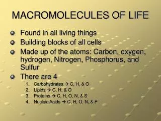
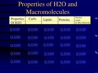
Macromolecules of life structure And properties • Nucleic acids • Proteins • Sugars • Lipids
Experiment by Griffith 1928 Pneumonia diplococcus Living bacteria of virulent strain Disease Living bacteria of non virulent strain Healthy mice Dead bacteria of virulent strain Heat attenuated Healthy mice Dead bacteria of virulent strain + Living non virulent bacteria Disease
Bacteria transformation in vitro Experiment by Avery 1943 Extraction of chemical cellular compounds from dead bacteria, Virulent bacteria strain Non virulent bacteria strain Mice got sick = extract from virulent bacteria transforms non virulent to virulent bacteria Enzymes digesting proteins (proteolytic) Non virulent bacteria strain Mice got sick = extract from Proteins are not transforming the factor Extract from virulent bacteria strain Extract after proteolytic digestion Enzymes digesting nucleic acids (nucleoliytc) Non virulent bacteria strain Mice are health = extract free of nucleic acids is not transforming bacteria Extract from virulent bacteria strain Extract after nucleolytic digestion DNA isolation Virulent bacteria strain Non virulent bacteria strain Mice got sick = DNA is the bacteria transforming factor DNA isolation from virulent bacteria
Alfred Harshey and Martha Chase – provided evidence that the DNA is the genetic material (1952) Virus separation following centifugation Labeling with 35S labeling with 32P isotope Homogenization in a mixer Bacteryophage proliferating in culture medium enriched with 35S and 32P has a coat labeled with 35S; core DNA labeled with 32P 35S Virus infecting bacteria 35S Protein coat in the supernatant 32P Bacteria containing DNA labeled with 32P isotope in the pelet. Virus replication Cell decay
Nucleic acids components(s u g a r s) ribose 2-deoxyribose
Nitrogen bases Pyrimidine (Py) Cytosine (C) Uracil (U) Thymine (T) Purines (Pu) Adenine (A) Guanine (G)
Nucleotides Purine nucleotides Guanine Adenosine Pyrimidine nucleotides Thymidine Cytosine
DNA molecule Structure Deoxyribonucleic acid (a fragment) 3’
Inventors of DNA structure - 1953 James Watson Francis Crick Maurice Wilkins Rosalind Franklin
Double stranded dna molecule structure 1. Two strands of the DNA form right-hand twisted double helix. 2. Anti-parallel nitrogen bases are linked to each other via hydrogen bonds accordingly to their complementarity: A/T and G/C. 3. The strands are anti-parallel. 4. There is 10 base pairs per single helix turn (360°). big grove small grove
Types of dnaSTRUcTURe Groves smaller bigger A-DNA Z-DNA B-DNA B-DNA A-DNA Z-DNA
RNA ribose 2-deoxyribose
Nitrogenbases in RNA Pyrimidine bases (Py) Cytosine (C) Uracil (U) Purinebases (Pu) Adenine (A) Guanine (G)
Ribonucleic acid 3’ 5’
proteins Peptide group Peptide unit is a rigid flat group of four atoms (N, H, C and O) standard bond lengths are denoted (in A)
proteins Peptide bond Peptide bond formation
STEReoisomers (enantiomers) • Mirror reflections (placement of 4 different asymetric carbon substitutes) • D- adopt as a standard D-glyceric aldehyde • L- adopt as a standard L-glyceric aldehyde • a standard L-glyceric aldehyde • The proteins consist of only L-amino acids • The optically active compounds Configurations of absolute isomers L and D of amino acids. R – a side chain. Isomers D and L are mirror reflections.
. Amino acids
Amino acids with hydrophobicalifatic side chain Alanine (Ala) Glycine (Gly) Vlanine (Vla) Leucine (leu) Isoleucine (Ile)
Amino acids hydrophobic with aromatic side chain Tryptofane (Trp) Thyrosine (Thr) Phenylalanine (Phe)
Hydrophobic amino acid Proline side chain contains cyclic structure
Amino acids with hydrophobic side chains containing sulfur Cysteine (Cys) Methionine (Met)
Amino acids with Aliphatic side chain containing hydroxyl group (hydrophilic chains ) Threonine (Thr) Serine (Ser)
Hydrophilic amino acids with basic side chains Arginine (Arg) Histidine (His) Lysine (Lys)
Hydrophilic amino acids with acidic side chains Aspargic acid (Asp) Glutamic acid (Glu)
Amide derivative of amino acids with acidic side chains Glutamine (Gln) Asparagine (Asn)
STRUCTURAL ORDERS OF POLYPEPTIDE CHAINS A primary order – an amino acid sequence. A protein consists of a polypeptide backbone with attached side chains. Each type of protein differs in its sequence and number of amino acids; therefore, it is the sequence of the chemically different side chains that makes each protein distinct. The two ends of a polypeptide chain are chemically different: the end carrying the free amino group (NH3+, also written NH2) is the amino terminus, or N-terminus, and that carrying the free carboxyl group (COO–, also written COOH) is the carboxyl terminus or C-terminus. The amino acid sequence of a protein is always presented in the N-to-C direction, reading from left to right.
STRUCTURAL ORDERS OF POLYPEPTIDE CHAINS Large numbers of hydrogen bonds form between adjacent regions of the folded polypeptide chain and help stabilize its three-dimensional shape. The protein depicted is a portion of the enzyme lysozyme, and the hydrogen bonds between the three possible pairs of partners have been differently colored, as indicated.
Secondary and tertiary order structures The regular conformation of the polypeptide backbone observed in the α helix and the β sheet (A, B, and C) The α helix. The N–H of every peptide bond is hydrogen-bonded to the C=O of a neighboring peptide bond located four peptide bonds away in the same chain. (D, E, and F) The β sheet. In this example, adjacent peptide chains run in opposite (antiparallel) directions. The individual polypeptide chains (strands) in a β sheet are held together by hydrogen-bonding between peptide bonds in different strands, and the amino acid side chains in each strand alternately project above and below the plane of the sheet. (A) and (D) show all the atoms in the polypeptide backbone, but the amino acid side chains are truncated and denoted by R. In contrast, (B) and (E) show the backbone atoms only, while (C) and (F) display the shorthand symbols that are used to represent the α helix and the β sheet in ribbon drawings of proteins.
Fourth order structure A protein formed from four domains In the Src protein shown, two of the domains form a protein kinase enzyme, while the SH2 and SH3 domains perform regulatory functions. (A) A ribbon model, with ATP substrate in red. (B) A spacing-filling model, with ATP substrate in red. Note that the site that binds ATP is positioned at the interface of the two domains that form the kinase. The detailed structure of the SH2 domain is illustrated in Panel 3-2 (pp. 138–139).
Protein Functions • Proteins functions might be but are not limited to: • Catalytic • Structural • Motility • Transporting • Homeostatic • Other
IZOMERs L and D – positioning of H and OH substitutes at the asymmetric carbon farthest from the aldehyde or ketone α and β –positioning of substituents at the ring mold sugar + and - - indicate the direction of light polarization: "+" - clockwise "-„ - left-hand L-glucose D-glucose D-glycer aldehyde L-glycer aldehyde β-D(+)-glucose (glucopyranose) α-D(+)-glucose (glucopyranose)
D-ketose family (1); D-erythrulose (2); D-ribulose (3a); D-xylulose (3b); D-psicose (4a); D-fructose (4b); D-sorbose (4c); D-tagatose (4d)
D-aldose family D-(+)-glyceraldehyde D-(−)-erythrose D-(−)-threose D-(−)-ribose; D-(−)-arabinose; D-(+)-xylose; D-(−)-lyxose D-(+)-allose; D-(+)-altrose; D-(+)-glucose; D-(+)-mannose; D-(−)-gulose; D-(−)-idose; D-(+)-galactose; D-(+)-talose
Pyranose ring forms • Five-membered rings are the most stable form of some carbohydrates. • For example D-fructose, a ketohexose, forms a stable five-membered ring. • Ribose, a common aldopentose, also forms a five-membered ring.
Pyranose ring forms • Ribose is important in biochemistry because it is the “sugar” part of the RNA bases such as cytidine. • Some sugars are missing –OH groups, and these are indicated by the number corresponding to the location of the missing –OH group, followed by the term “deoxy” meaning “without oxygen”. • 2-Deoxy-D-ribose is the “sugar” on DNA bases, for example the base 2’-deoxycytidine.
Furanose Ring forms Cycliztion: • hemiacetalbond formation between the-OH group at the carbon to the carbonyl group the fourth and the first carbon atom • hemiketalbond formation between the carbonyl group at the second carbon atom and the group-OH at the fifth carbon furan 14
Functions of sugars • Source of energy • Storage of energetic material – glicogen, starch • Building material – components of cell wall in plant cells
Fatty acids • Saturated fatty acids: CH3(CH2)nCOOH n from 12 to 24
Fatty acidsNomenclature of fatty acids • Systematic names derive from Greek numerical • Location od double bond is marked by symbolD,and carbon numbers participating in double bonds formation, are counted from carboxyl group • letternorwindicate position of double bond, counting from methyl group. w Kwastłuszczowy z wiązaniemcisD-9
Fatty acids Unsaturated Fatty acids