Download
1 / 35
360 likes | 636 Views
Basal Ganglia Principles of neural sciences 5 th ed. The human brain: an introduction to its functional anatomy 6 th ed. 林 永 煬 國立陽明大學 腦科學研究所. Basal ganglia and surrounding structures, as seen in an axial section. Basal ganglia and surrounding structures, as seen in coronal sections.

E N D
Basal Ganglia Principles of neural sciences 5th ed. The human brain: an introduction to its functional anatomy 6th ed. 林 永 煬 國立陽明大學 腦科學研究所
Basal ganglia and surrounding structures, as seen in an axial section
Basal ganglia and surrounding structures, as seen in coronal sections
The compact (SNc) and reticular (SNr) parts of the substantia nigra
Three-dimensional reconstruction of the striatum and globus pallidus inside a translucent CNS
Striopallidal fibers: from putamen to globus pallidus (pallidum) Corticostriate fibers: from cerebral cortex to putamen, caudate nucleus, or nucleus accumbens Pallidothalamic fibers: from globus pallidus to thalamus Nigrostriatal fibers: from substantia nigra to striatum Nigroreticular fibers: from substantia nigra to reticular formation Thalamostriate fibers: from intralaminar nuclei to striatum
Basal ganglia circuitry Corticostriate inputs: Afferents from cortex to striatum and subthalamic nucleus Excitatory (glutamate) connections Output projections: Efferents leaving from globus pallidus (GPi) and substantia nigra (SNr) to thalamus Inhibitory (r-aminobutyric acid [GABA]) connections Thalamocortical projections: Excitatory (glutamate) connections
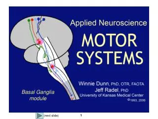
Cortical functions related to basal ganglia circuitry Movement Cognition Emotion Motivation
Cortical functions related to basal ganglia circuitry Motor functions: sensorimotor cortex putamen GP thalamus motor/premotor/supplementary motor areas Cognitive functions: association cortex caudate nucleus GPi-SNr thalamus prefrontal areas Emotion/motivation: amygdala/hippocampus/limbic cortex ventral striatum (nucleus accumbens) ventral pallidum thalamus (dorsomedial nucleus) temporal/ hippocampus/limbic cortex
Medial (A) and lateral (B) views of the left striatum showing the somatotopic representation of body parts
Chemical compartmentalization of the striatum * * CN IC P A P=putamen, CN=caudate nucleus, IC=internal capsule, A=nucleus accumbens Acetylcholinesterase (AChE)-rich background (matrix) with embedded AChE-poor regions (striosomes *) Caudate nucleus stained for enkephalin Caudate nucleus stained for AChE High enkephalin levels are found in the peripheries of striosomes.
Case presentation A 25-year-old woman with dramatic change in cognition, motivation, and self-care.
Contrast-enhanced computed tomography scans of a 25-year-old woman with dramatic change in cognition, motivation, and self-care. Acute stage 8 months later
Major connections of the external segment of the globus pallidus (GPe)
Afferents to GPi and SNr Efferents from GPi and SNr GPi and SNr receive inhibitory inputs from striatum, and excitatory inputs from STN. GPi and SNr provide output from basal ganglia.
Major connections of STN STN provides a powerful excitatory input to GPi and SNr. Major inputs to STN arise not only from GP but also from cerebral cortex. Different sectors of STN deal with motor, cognitive, affective functions.
Abnormalities of movement due to basal ganglia disorders Hyperkinetic disorders: tremor, chorea, athetosis, ballismus, hemiballismus Disturbance of muscle tone: dystonia Huntington’s disease Parkinson’s disease
Hemiballismus Video show s/s: wild flailing movements of one arm and leg Lesion location in contralateral STN Most often seen in older people with a stroke involving a ganglionic branch of posterior cerebral artery
Contrast-enhanced mass in left STN of a 65 y/o male HIV-positive patient with unintentional, forceful flinging movements of right limbs. Longitudinal slice of a normal brain
Huntington’s disease s/s: involuntary choreiform movements, alterations of mood or cognitive function dementia and personality change Symptom onset at age of 30 – 50 years Inherited in an autosomal dominant pattern, with a defective gene at short arm of chromosome 4 Pathomechanism: striatal (esp. caudate nucleus) and cortical degeneration Video show
MRI scan in a 29 y/o man with Huntington's disease MRI scan in a normal individual Dorsal-ventral degeneration gradient (red > blue)
Parkinson’s disease s/s: tremor rigidity (increased muscle tone) slow movements (bradykinesia, hypokinesia) stooped posture Pathomechanism: degeneration of substantia nigra (esp. SNc, pigmented nigral cells)
The midbrain of a patient with Parkinson's disease, showing loss of pigmentation in the compact part of the substantia nigra (*)
A model for movement control by basal ganglia (through excitatory (green) and inhibitory (red) interactions) Indirect pathway Direct pathway Loss of dopamine neurons from SNc causes a reduced thalamic and cortical output. (e.g. PD) Loss of excitatory STN projections (e.g. hemiballismus) Dopamine excites direct pathway, but inhibits indirect pathway.
Increased blood flow in the supplementary motor area (S) and premotor cortex (P) of Parkinson's disease patients during movement following treatment with levo-dopa
Parkinson’s disease following unilateral pallidotomy Improvement in bradykinesia and rigidity Increase in cerebral blood flow on PET