Download
1 / 7
80 likes | 278 Views
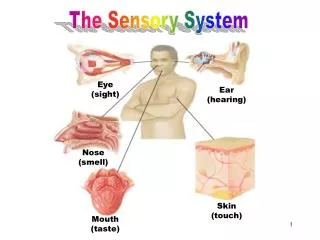
SENSORY SYSTEM. STRUCTURE OF THE EYE. EYE. 1 ” in diameter Protected by orbital cavity, eyebrows, eyelashes, eyelids Lacrimal glands Tears empty into nasal cavity Conjunctiva Thin membrane lines eyelids Wall of eye made up of three coats Sclera Choroid Retina

E N D
SENSORY SYSTEM STRUCTURE OF THE EYE
EYE • 1” in diameter • Protected by orbital cavity, eyebrows, eyelashes, eyelids • Lacrimal glands • Tears empty into nasal cavity • Conjunctiva • Thin membrane lines eyelids • Wall of eye made up of three coats • Sclera • Choroid • Retina • Pages 189 and 190 figures 1 & 2
SCLERA • Outer layer • White of the eye • Tough coating, helps maintain shape of eye • Muscles responsible for moving eye attached to sclera: • Extrinsic muscles • See figure 10-3 and Table 10-1 pg 191
CORNEA • Front center of sclera (clear part) no blood vessels • Transparent so light rays can pass through • Considered the “window” of the eye
CHOROID COAT • Middle layer, contains blood vessels • Opening in front is pupil • Colored, muscular layer surrounding pupil is the iris • Intrinsic muscles • Change size of iris to control amount of light entering through pupil
LENS • Crystalline structure located behind iris and pupil • Elastic, disc-shaped, biconvex • Job is to focus images on the retina • Situated between the anterior and posterior chambers • Anterior Chamber – filled with aqueous humor (watery fluid) and constantly replenished by blood vessels behind the iris • Posterior Chamber – filled with vitreous humor (transparent jelly-like substance) • Both help maintain the eyeball’s shape
RETINA • Innermost layer • Light rays focus image on retina • Image travels to the cerebral cortex via optic nerve • Rods • Sensitive to dim light • Cones • Sensitive to bright light and color • Optic disc • On retina, known as blind spot, nerve fibers that form optic nerve • See figures 10-5 and 10-6 pgs. 192-193